millirem – kilorem. Equivalent doses measured in industry and medicine often have usually lower doses than one rem, and the following multiples are often used:1 mrem (millirem) = 1E-3 rem and 1 krem (kilorem) = 1E3 rem
In radiation protection, the rem (an abbreviation for Roentgen Equivalent Man) is the non-SI unit of the equivalent dose, which is used predominantly in the USA. The rem represents the equivalent biological effect of the deposit of one hundred ergs (one rad) of gamma rays energy in a kilogram of human tissue. The rem is not derived from the unit of exposure, the roentgen. The acronym is now a misleading historical artifact, since 1 roentgen actually deposits about 0.96 rem in soft biological tissue, when all weighting factors equal unity.
As was written, the rem is used for radiation dose quantities such as equivalent dose and effective dose. Equivalent dose (symbol HT) is a dose quantity calculated for individual organs (index T – tissue). Equivalent dose is based on the absorbed dose to an organ, adjusted to account for the effectiveness of the type of radiation. The SI unit of HT is the sievert (Sv) or but rem (roentgen equivalent man) is still commonly used (1 Sv = 100 rem).
One rem is also a large amount of equivalent dose. A person who has absorbed a whole body dose of 1 rem has absorbed one hundred ergs of energy in each kg of body tissue (in case of gamma rays).
Equivalent doses measured in industry and medicine often have usually lower doses than one rem, and the following multiples are often used:
1 mrem (millirem) = 1E-3 rem
1 krem (kilorem) = 1E3 rem
Conversions from the SI units to other units are as follows:
1 Sv = 100 rem
1 mSv = 100 mrem
Examples of Doses in millirems
We must note that radiation is all around us. In, around, and above the world we live in. It is a natural energy force that surrounds us. It is a part of our natural world that has been here since the birth of our planet. In the following points we try to express enormous ranges of radiation exposure, which can be obtained from various sources.
0.005 mrem – Sleeping next to someone
0.009 mrem – Living within 30 miles of a nuclear power plant for a year
0.01 mrem – Eating one banana
0.03 mrem – Living within 50 miles of a coal power plant for a year
1 mrem – Average daily dose received from natural background
2 mrem – Chest X-ray
4 mrem – A 5-hour airplane flight
60 mrem – mammogram
100 mrem – Dose limit for individual members of the public, total effective dose per annum
365 mrem – Average yearly dose received from natural background
580 mrem – Chest CT scan
1 000 mrem – Average yearly dose received from natural background in Ramsar, Iran
2 000 mrem – single full-body CT scan
17 500 mrem – Annual dose from natural radiation on a monazite beach near Guarapari, Brazil.
500 000 mrem – Dose that kills a human with a 50% risk within 30 days (LD50/30), if the dose is received over a very short duration.
Robert Reed Burn, Introduction to Nuclear Reactor Operation, 1988.
U.S. Department of Energy, Nuclear Physics and Reactor Theory. DOE Fundamentals Handbook, Volume 1 and 2. January 1993.
Paul Reuss, Neutron Physics. EDP Sciences, 2008. ISBN: 978-2759800414.
See also:
REM
We hope, this article, millirem – kilorem – multiples of rem, helps you. If so, give us a like in the sidebar. Main purpose of this website is to help the public to learn some interesting and important information about radiation and dosimeters.
Intensity of radiation is a key factor, which determines health effects from being exposed to any radiation. The equivalent dose rate is the rate at which an equivalent dose is received. Radiation Dosimetry
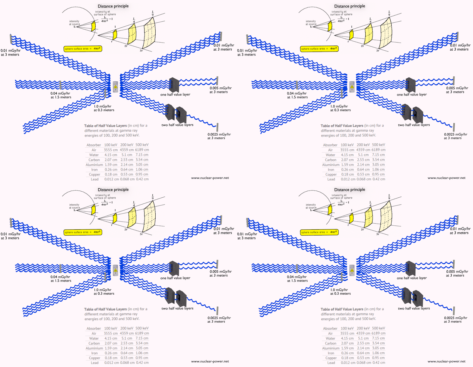
Principles of Radiation Protection – Time, Distance, Shielding
Intensity of radiation is a key factor, which determines health effects from being exposed to any radiation. It is similar as being exposed to heat radiation from a fire (in fact, it is also transferred by photons). If you are too close to a fire, the intensity of thermal radiation is high and you can get burned. If you are at the right distance, you can withstand there without any problems and moreover it is comfortable. If you are too far from heat source, the insufficiency of heat can also hurt you. This analogy, in a certain sense, can be applied to radiation also from ionizing radiation sources.
In short, to get burned (deterministic effects and demonstrable stochastic effects) by ionizing radiation, you must be exposed to really high amount of radiation. But almost everytime, we are talking about so called low doses. As was written, today the protection system is based on the LNT-hypothesis, which is a conservative model used in radiation protection to estimate the health effects from small radiation doses. This model is excellent for setting up a protection system for all use of ionizing radiation. This model assumes, that there is no threshold point and risk increases linearly with a dose, i.e. the LNT model implies that there is no safe dose of ionizing radiation. If this linear model is correct, then natural background radiation is the most hazardous source of radiation to general public health, followed by medical imaging as a close second. It must be added, the research during the last two decades is very interesting and show that small doses of radiation given at a low dose rate stimulate the defense mechanisms. Therefore the LNT model is not universally accepted with some proposing an adaptive dose–response relationship where low doses are protective and high doses are detrimental. Many studies have contradicted the LNT model and many of these have shown adaptive response to low dose radiation resulting in reduced mutations and cancers. On the other hand, it is very important, to what type of radiation is a person exposed.
Equivalent Dose Rate
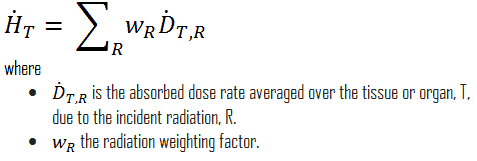
The equivalent dose rate is the rate at which an equivalent dose is received. It is a measure of radiation dose intensity (or strength). The equivalent dose rate is therefore defined as:
In conventional units, it is measured in mSv/sec, Sv/hr, mrem/sec or rem/hr. Since the amount of radiation exposure depends directly (linearly) on the time people spend near the source of radiation, the absorbed dose is equal to the strength of the radiation field (dose rate) multiplied by the length of time spent in that field. The example above indicates a person could expect to receive a dose of 25 millirems by staying in a 50 millirems/hour field for thirty minutes.
Sievert and Gray
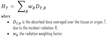
For radiation protection purposes, the absorbed dose is averaged over an organ or tissue, T, and this absorbed dose average is weighted for the radiation quality in terms of the radiation weighting factor, wR, for the type and energy of radiation incident on the body. The radiation weighting factor is a dimensionless factor used to determine the equivalent dose from the absorbed dose averaged over a tissue or organ and is based on the type of radiation absorbed. The resulting weighted dose was designated as the organ- or tissue equivalent dose:
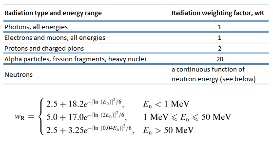
Table of radiation weighting factors. Source: ICRP Publ. 103: The 2007 Recommendations of the International Commission on Radiological Protection
An equivalent dose of one Sievert represents that quantity of radiation dose that is equivalent, in terms of specified biological damage, to one gray of X-rays or gamma rays. A dose of one Sv caused by gamma radiation is equivalent to an energy deposition of one joule in a kilogram of a tissue. That means one sievert is equivalent to one gray of gamma rays deposited in certain tissue. On the other hand, similar biological damage (one sievert) can be caused only by 1/20 gray of alpha radiation (due to high wR of alpha radiation). Therefore, the sievert is not a physical dose unit. For example, an absorbed dose of 1 Gy by alpha particles will lead to an equivalent dose of 20 Sv. This may seem to be a paradox. It implies that the energy of the incident radiation field in joules has increased by a factor of 20, thereby violating the laws of Conservation of energy. However, this is not the case. Sievert is derived from the physical quantity absorbed dose, but also takes into account the biological effectiveness of the radiation, which is dependent on the radiation type and energy. The radiation weighting factor causes that the sievert cannot be a physical unit.
One sievert is a large amount of equivalent dose. A person who has absorbed a whole body dose of 1 Sv has absorbed one joule of energy in each kg of body tissue (in case of gamma rays).
Equivalent doses measured in industry and medicine often have usually lower doses than one sievert, and the following multiples are often used:
1 mSv (millisievert) = 1E-3 Sv
1 µSv (microsievert) = 1E-6 Sv
Conversions from the SI units to other units are as follows:
1 Sv = 100 rem
1 mSv = 100 mrem
Examples of Doses in Sieverts
We must note that radiation is all around us. In, around, and above the world we live in. It is a natural energy force that surrounds us. It is a part of our natural world that has been here since the birth of our planet. In the following points we try to express enormous ranges of radiation exposure, which can be obtained from various sources.
0.05 µSv – Sleeping next to someone
0.09 µSv – Living within 30 miles of a nuclear power plant for a year
0.1 µSv – Eating one banana
0.3 µSv – Living within 50 miles of a coal power plant for a year
10 µSv – Average daily dose received from natural background
20 µSv – Chest X-ray
40 µSv – A 5-hour airplane flight
600 µSv – mammogram
1 000 µSv – Dose limit for individual members of the public, total effective dose per annum
3 650 µSv – Average yearly dose received from natural background
5 800 µSv – Chest CT scan
10 000 µSv – Average yearly dose received from natural background in Ramsar, Iran
20 000 µSv – single full-body CT scan
175 000 µSv – Annual dose from natural radiation on a monazite beach near Guarapari, Brazil.
5 000 000 µSv – Dose that kills a human with a 50% risk within 30 days (LD50/30), if the dose is received over a very short duration.
Robert Reed Burn, Introduction to Nuclear Reactor Operation, 1988.
U.S. Department of Energy, Nuclear Physics and Reactor Theory. DOE Fundamentals Handbook, Volume 1 and 2. January 1993.
Paul Reuss, Neutron Physics. EDP Sciences, 2008. ISBN: 978-2759800414.
See also:
Radiation Exposure
We hope, this article, Intensity of Radiation – Dose and Dose Rate, helps you. If so, give us a like in the sidebar. Main purpose of this website is to help the public to learn some interesting and important information about radiation and dosimeters.
Radiation Protection Principles – Time – Distance – Shielding. For external exposure, we can use some of radiation protection principles (time, distance, shielding). Radiation Dosimetry
ionizing radiation – hazard symbol
Radiation protection is the science and practice of protecting people and the environment from the harmful effects of ionizing radiation. The International Atomic Energy Agency (IAEA) defines radiation protection as:
“The protection of people from harmful effects of exposure to ionizing radiation, and the means for achieving this”
It is a serious topic not only in nuclear power plants, but also in industry or in medical centres. According to the IAEA, radiation protection can be divided into three groups:
occupational radiation protection, which is the protection of workers in situations where their exposure is directly related to or required by their work
medical radiation protection, which is the protection of patients exposed to radiation as part of their diagnosis or treatment
public radiation protection, which is the protection of individual members of the public and of the population in general
According to the ICRP (Publication 103), the System of Radiological Protection is based on the following three principles:
Justification. “Any decision that alters the radiation exposure situation should do more good than harm.”
Optimisation of Protection. “Doses should all be kept as low as reasonably achievable, taking into account economic and societal factors.” (known as ALARA or ALARP)
Dose Limitation. “The total dose to any individual … should not exceed the appropriate limits.”
See also: ICRP, 2007. The 2007 Recommendations of the International Commission on Radiological Protection. ICRP Publication 103. Ann. ICRP 37 (2-4).
Radiation Protection Principles – Time – Distance – Shielding
External exposure is radiation that comes from outside our body and interacts with us. In this case, we analyze predominantly exposure from gamma rays, since alpha and beta particles, in general, constitute no external exposure hazard because the particles generally do not pass through skin. The source of radiation can be, for example, a piece of equipment that produces the radiation like a container with a radioactive materials, or like an x-ray machine. In radiation protection there are three ways how to protect people from identified external radiation sources:
Principles of Radiation Protection – Time, Distance, Shielding
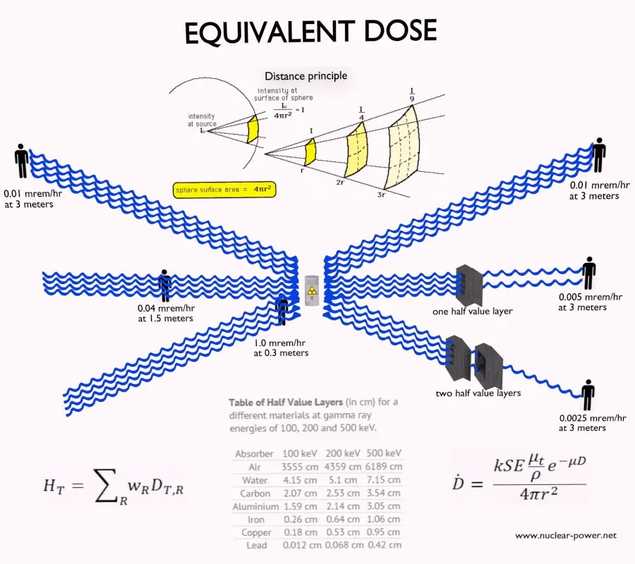
Limiting Time. The amount of radiation exposure depends directly (linearly) on the time people spend near the source of radiation. The dose can be reduced by limiting exposure time.
Distance. The amount of radiation exposure depends on the distance from the source of radiation. Similarly to a heat from a fire, if you are too close, the intensity of heat radiation is high and you can get burned. If you are at the right distance, you can withstand there without any problems and moreover it is comfortable. If you are too far from heat source, the insufficiency of heat can also hurt you. This analogy, in a certain sense, can be applied to radiation also from radiation sources.
Shielding. Finally, if the source is too intensive and time or distance do not provide sufficient radiation protection, the shielding must be used. Radiation shielding usually consist of barriers of lead, concrete or water. There are many many materials, which can be used for radiation shielding, but there are many many situations in radiation protection. It highly depends on the type of radiation to be shielded, its energy and many other parametres. For example, even depleted uranium can be used as a good protection from gamma radiation, but on the other hand uranium is absolutely inappropriate shielding of neutron radiation.
Robert Reed Burn, Introduction to Nuclear Reactor Operation, 1988.
U.S. Department of Energy, Nuclear Physics and Reactor Theory. DOE Fundamentals Handbook, Volume 1 and 2. January 1993.
Paul Reuss, Neutron Physics. EDP Sciences, 2008. ISBN: 978-2759800414.
See also:
Radiation Protection
We hope, this article, Radiation Protection Principle – Time – Distance – Shielding, helps you. If so, give us a like in the sidebar. Main purpose of this website is to help the public to learn some interesting and important information about radiation and dosimeters.
For external exposure, we cannot use some of radiation protection principles (time, distance, shielding). External contamination means that radioactive material has been deposited on surfaces (such as clothes, shoes). Radiation Dosimetry
External Dose Uptake
External exposure is radiation that comes from outside our body and interacts with us. In this case, we analyze predominantly exposure from gamma rays, since alpha and beta particles, in general, constitute no external exposure hazard because the particles generally do not pass through skin. The source of radiation can be, for example, a piece of equipment that produces the radiation like a container with a radioactive materials, or like an x-ray machine. In radiation protection there are three ways how to protect people from identified external radiation sources:
Principles of Radiation Protection – Time, Distance, Shielding
Limiting Time. The amount of radiation exposure depends directly (linearly) on the time people spend near the source of radiation. The dose can be reduced by limiting exposure time.
Distance. The amount of radiation exposure depends on the distance from the source of radiation. Similarly to a heat from a fire, if you are too close, the intensity of heat radiation is high and you can get burned. If you are at the right distance, you can withstand there without any problems and moreover it is comfortable. If you are too far from heat source, the insufficiency of heat can also hurt you. This analogy, in a certain sense, can be applied to radiation also from radiation sources.
Shielding. Finally, if the source is too intensive and time or distance do not provide sufficient radiation protection, the shielding must be used. Radiation shielding usually consist of barriers of lead, concrete or water. There are many many materials, which can be used for radiation shielding, but there are many many situations in radiation protection. It highly depends on the type of radiation to be shielded, its energy and many other parametres. For example, even depleted uranium can be used as a good protection from gamma radiation, but on the other hand uranium is absolutely inappropriate shielding of neutron radiation.
As was written, it is crucial, whether we are exposed to radiation from external sources or from internal sources. This is similar as for another dangerous substances. Internal exposure is more dangerous than external exposure, since we are carrying the source of radiation inside our bodies and we cannot use any of radiation protection principles (time, distance, shielding).
ionizing radiation – hazard symbol
Radiation Exposure
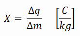
In general, radiation exposure is a measure of the ionization of air due to ionizing radiation from high-energy photons(i.e. X-rays and gamma rays). Radiation exposure is defined as the sum of electrical charges (∆q) on all the ions of one sign produced in air when all the electrons, liberated by photons in a volume of air whose mass is ∆m, are completely stopped in air.
Radiation exposure is given the symbol X. The SI unit of radiation exposure is the coulomb per kilogram (C/kg), but in practice, the roentgen is used. The roentgen, abbreviated R, is the unit of radiation exposure. In the original definition 1 R means the amount of X-rays or γ-radiation that is required to liberate positive and negative charges of one electrostatic unit of charge (esu) in 1 cm³ of dry air at standard temperature and pressure (STP).
Absorbed and Equivalent Dose
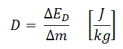
In radiation protection, the sievert is a derived unit of equivalent dose and effective dose. The sievert represents the equivalent biological effect of the deposit of a joule of gamma rays energy in a kilogram of human tissue. Absorbed dose is defined as the amount of energy deposited by ionizing radiation in a substance. Absorbed dose is given the symbol D. The absorbed dose is usually measured in a unit called the gray (Gy), which is derived from the SI system. The non-SI unit rad is sometimes also used, predominantly in the USA.
For radiation protection purposes, the absorbed dose is averaged over an organ or tissue, T, and this absorbed dose average is weighted for the radiation quality in terms of the radiation weighting factor, wR, for the type and energy of radiation incident on the body. The radiation weighting factor is a dimensionless factor used to determine the equivalent dose from the absorbed dose averaged over a tissue or organ and is based on the type of radiation absorbed. The resulting weighted dose was designated as the organ- or tissue equivalent dose:
Table of radiation weighting factors. Source: ICRP Publ. 103: The 2007 Recommendations of the International Commission on Radiological Protection
An equivalent dose of one Sievert represents that quantity of radiation dose that is equivalent, in terms of specified biological damage, to one gray of X-rays or gamma rays. A dose of one Svcaused by gamma radiation is equivalent to an energy deposition of one joule in a kilogram of a tissue. That means one sievert is equivalent to one gray of gamma rays deposited in certain tissue. On the other hand, similar biological damage (one sievert) can be caused only by 1/20 gray of alpha radiation (due to high wR of alpha radiation). Therefore, the sievert is not a physical dose unit. For example, an absorbed dose of 1 Gy by alpha particles will lead to an equivalent dose of 20 Sv. This may seem to be a paradox. It implies that the energy of the incident radiation field in joules has increased by a factor of 20, thereby violating the laws of Conservation of energy. However, this is not the case. Sievert is derived from the physical quantity absorbed dose, but also takes into account the biological effectiveness of the radiation, which is dependent on the radiation type and energy. The radiation weighting factor causes that the sievert cannot be a physical unit.
Contamination versus Radiation
Radioactive contamination consist of radioactive material, that generate ionizing radiation. It is the source of radiation, not radiation itself. Anytime that radioactive material is not in a sealed radioactive source container and might be spread onto other objects, radioactive contamination is a possibility. Radioactive contamination may be characterized by following points:
Radioactive contamination consist of radioactive material (contaminants), that may be solid, liquid or gaseous. Large contaminants can be even visible, but you cannot see radiation produced.
When released, contaminants can be spread by air, water or just by mechanical contact.
We cannot shield contamination.
We can mitigate contamination by protecting integrity of barriers (source container, fuel cladding, reactor vessel, containment building)
Since contaminants interact chemically, they may be contained within objects such as the human body.
We can rid of contamination by many mechanical, chemical (decontaminate surfaces), or biological processes (biological half-life).
It is of the highest importance, which material is the radioactive contaminant (half-life, mode of decay, energy).
Ionizing radiation is formed by high-energy particles (photons, electrons, etc.), that can penetrate matter and ionize (to form ion by losing electrons) target atoms to form ions. Radiation exposure is the consequence of the presence nearby the source of radiation. Radiation exposure as a quantity is defined as a measure of the ionization of material due to ionizing radiation. The danger of ionizing radiation lies in the fact that the radiation is invisible and not directly detectable by human senses. People can neither see nor feel radiation, yet it deposits energy to the molecules of the body. The energy is transferred in small quantities for each interaction between radiation and a molecule and there are usually many such interactions. Unlike radioactive contamination, radiation may be characterized by following points:
Radiation consist consist of high-energy particles that can penetrate matter and ionize (to form ion by losing electrons) target atoms. Radiation is invisible, and not directly detectable by human senses. It must be noted, beta radiation is indirectly visible due to cherenkov radiation.
Unlike contamination, radiation cannot be spread by any medium. It travels through materials until it loses its energy. We can shield radiation (e.g. by standing around the corner).
Exposure to ionizing does not necessary mean, that the object becomes radioactive (except very rare neutron radiation).
Radiation can penetrate barriers, but sufficiently thick barrier can minimize all effects.
Unlike contaminants, radiation cannot interact chemically with matter and cannot be bound inside body.
It is not important, which material is the source of certain radiation. Only type of radiation and energy matters.
External Contamination
External contaminationmeans that radioactive material has been deposited on surfaces (such as walls, floors). It may be loosely deposited, much like ordinary dust, or it may be quite firmly fixed by chemical reaction. This distinction is important, and we classify surface contamination on the basis of how easily it can be removed:
Free Contamination. In the case of free contamination (or loose contamination), the radioactive material can be spread. This is surface contamination that can easily be removed with simple decontamination methods. For example, if dust particles containing various radioisotopes land on the person’s skin or garments, we can clean it up or remove clothes. Once a person has been decontaminated, all of the particulate radioactivity sources are eliminated, and the individual is no longer contaminated. Free contamination is also a more serious hazard than fixed contamination, because dust particles can become airborne and they can be easily ingested. This leads to an internal exposure by radioactive contaminants. Although almost all contaminants are beta radioactive with accompanying gamma emission, but there is also the possibility of alpha contamination in any nuclear fuel handling areas.
Fixed Contamination. In the case of fixed contamination, the radioactive material cannot be spread, since it is chemically or mechanically bound to structures. It cannot be removed by normal cleaning methods. Fixed contamination is a less serious hazard than free contamination, it cannot be re-suspended or transferred to skin. Therefore the hazard is usually an external one only. On the other hand, it depends on the level of contamination. Although almost all contaminants are beta radioactive with accompanying gamma emission, but there is also the possibility of alpha contamination in any nuclear fuel handling areas. Unless the level of contamination is very severe, the gamma radiation dose rate will be small and external exposure will be significant only in contact with, or very close to, the contaminated surfaces. Since beta particles are less penetrating than gamma rays, the beta dose rate can be high only at contact. A value of 1 mSv/h at contact for a contamination level of 400 – 500 Bq/cm2 is fairly representative.
Robert Reed Burn, Introduction to Nuclear Reactor Operation, 1988.
U.S. Department of Energy, Nuclear Physics and Reactor Theory. DOE Fundamentals Handbook, Volume 1 and 2. January 1993.
Paul Reuss, Neutron Physics. EDP Sciences, 2008. ISBN: 978-2759800414.
See also:
Radiation Protection
We hope, this article, External Exposure – External Contamination, helps you. If so, give us a like in the sidebar. Main purpose of this website is to help the public to learn some interesting and important information about radiation and dosimeters.
If the source of radiation is inside our body, we say, it is internal exposure. Protection againts internal contamination is key for radiation protection. Radiation Dosimetry
ionizing radiation – hazard symbol
If the source of radiation is inside our body, we say, it is internal exposure. The intake of radioactive material can occur through various pathways such as ingestion of radioactive contamination in food or liquids. Protection from internal exposure is more complicated. Most radionuclides will give you much more radiation dose if they can somehow enter your body, than they would if they remained outside.
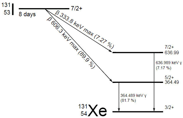
As was written, it is crucial, whether we are exposed to radiation from external sources or from internal sources. This is similar as for another dangerous substances. Internal exposure is more dangerous than external exposure, since we are carrying the source of radiation inside our bodies and we cannot use any of radiation protection principles (time, distance, shielding). The intake of radioactive material can occur through various pathways such as ingestion of radioactive contamination in food or liquids, inhalation of radioactive gases, or through intact or wounded skin. On this place, we have to distinguish between radiation and contamination. Radioactive contamination consist of radioactive material, that generate ionizing radiation. It is the source of radiation, not radiation itself. Anytime that radioactive material is not in a sealed radioactive source container and might be spread onto other objects, radioactive contamination is a possibility. For example, radioiodine, iodine-131, is an important radioisotope of iodine. Radioiodine plays a major role as a radioactive isotope present in nuclear fission products, and it is a major contributor to the health hazards when released into the atmosphere during an accident. Iodine-131 has a half-life of 8.02 days. The target tissue for radioiodine exposure is the thyroid gland. The external beta and gamma dose from radioiodine present in the air is quite negligible when compared to the committed dose to the thyroid that would result from breathing this air.
Internal Dose Uptake
If the source of radiation is inside our body, we say, it is internal exposure. The intake of radioactive material can occur through various pathways such as ingestion of radioactive contamination in food or liquids, inhalation of radioactive gases, or through intact or wounded skin. Most radionuclides will give you much more radiation dose if they can somehow enter your body, than they would if they remained outside. For internal doses, we first should distinguish between intake and uptake. Intake means what a person takes in. Uptake means what a person keeps.
When a radioactive compound enters the body, the activity will decrease with time, due both to radioactive decay and to biological clearance. The decrease varies from one radioactive compound to another. For this purpose, the biological half-life is defined in radiation protection.
The biological half-life is the time taken for the amount of a particular element in the body to decrease to half of its initial value due to elimination by biological processes alone, when the rate of removal is roughly exponential. The biological half-life depends on the rate at which the body normally uses a particular compound of an element. Radioactive isotopes that were ingested or taken in through other pathways will gradually be removed from the body via bowels, kidneys, respiration and perspiration. This means that a radioactive substance can be expelled before it has had the chance to decay.
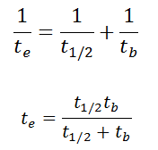
As a result, the biological half-life significantly influences the effective half-life and the overall dose from internal contamination. If a radioactive compound with radioactive half-life (t1/2) is cleared from the body with a biological half-life tb, the effective half-life (te) is given by the expression:
As can be seen, the biological mechanisms always decreases the overall dose from internal contamination. Moreover, if t1/2 is large in comparison to tb, the effective half-life is approximately the same as tb.
For example, tritium has the biological half-life about 10 days, while the radioactive half-life is about 12 years. On the other hand, radionuclides with very short radioactive half-lives have also very short effective half-lives. These radionuclides will deliver, for all practical purposes, the total radiation dose within the first few days or weeks after intake.
For tritium, the annual limit on intake (ALI) is 1 x 10⁹ Bq. If you take in 1 x 10⁹ Bq of tritium, you will receive a whole-body dose of 20 mSv. The committed effective dose, E(t), is therefore 20 mSv. It does not depend whether a person intakes this amount of activity in a short time or in a long time. In every case, this person gets the same whole-body dose of 20 mSv.
Contamination versus Radiation
Radioactive contamination consist of radioactive material, that generate ionizing radiation. It is the source of radiation, not radiation itself. Anytime that radioactive material is not in a sealed radioactive source container and might be spread onto other objects, radioactive contamination is a possibility. Radioactive contamination may be characterized by following points:
Radioactive contamination consist of radioactive material (contaminants), that may be solid, liquid or gaseous. Large contaminants can be even visible, but you cannot see radiation produced.
When released, contaminants can be spread by air, water or just by mechanical contact.
We cannot shield contamination.
We can mitigate contamination by protecting integrity of barriers (source container, fuel cladding, reactor vessel, containment building)
Since contaminants interact chemically, they may be contained within objects such as the human body.
We can rid of contamination by many mechanical, chemical (decontaminate surfaces), or biological processes (biological half-life).
It is of the highest importance, which material is the radioactive contaminant (half-life, mode of decay, energy).
Ionizing radiation is formed by high-energy particles (photons, electrons, etc.), that can penetrate matter and ionize (to form ion by losing electrons) target atoms to form ions. Radiation exposure is the consequence of the presence nearby the source of radiation. Radiation exposure as a quantity is defined as a measure of the ionization of material due to ionizing radiation. The danger of ionizing radiation lies in the fact that the radiation is invisible and not directly detectable by human senses. People can neither see nor feel radiation, yet it deposits energy to the molecules of the body. The energy is transferred in small quantities for each interaction between radiation and a molecule and there are usually many such interactions. Unlike radioactive contamination, radiation may be characterized by following points:
Radiation consist consist of high-energy particles that can penetrate matter and ionize (to form ion by losing electrons) target atoms. Radiation is invisible, and not directly detectable by human senses. It must be noted, beta radiation is indirectly visible due to cherenkov radiation.
Unlike contamination, radiation cannot be spread by any medium. It travels through materials until it loses its energy. We can shield radiation (e.g. by standing around the corner).
Exposure to ionizing does not necessary mean, that the object becomes radioactive (except very rare neutron radiation).
Radiation can penetrate barriers, but sufficiently thick barrier can minimize all effects.
Unlike contaminants, radiation cannot interact chemically with matter and cannot be bound inside body.
It is not important, which material is the source of certain radiation. Only type of radiation and energy matters.
Airborne Contamination
Airborne contamination is of particular importance in nuclear power plants, where it must be monitored. Contaminants can become airborne especially during reactor top head remove, reactor refueling, and during manipulations within spent fuel pool. The air can be contaminated with radioactive isotopes especially in particulate form, which poses a particular inhalation hazard. This contamination consists of various fission and activation products that enter the air in gaseous, vapour or particulate form. There are four types of airborne contamination in nuclear power plants, namely:
Particulates. Particulate activity is an internal hazard, because it can be inhaled. Transportable particulate material taken into the respiratory system will enter the blood stream and be carried to all parts of the body. Non-transportable particulates will stay in the lungs with a certain biological half-life. For example, Sr-90, Ra-226 and Pu-239 are radionuclides known as bone-seeking radionuclides. These radionuclides have long biological half-lives and are serious internal hazards. Once deposited in bone, they remain there essentially unchanged in amount during the lifetime of the individual. The continued action of the emitted alpha particles can cause significant injury: over many years they deposit all their energy in a tiny volume of tissue, because the range of the alpha particles is very short.
Noble gases. Radioactive noble gases, such as xenon-133, xenon-135 and krypton-85 are present in reactor coolant especially when fuel leakages are present. As they appear in coolant, they become airborne and they can be inhaled. They are exhaled right after they are inhaled, because the body does not react chemically with them. If workers are working in a noble gas cloud, the external dose they will receive is about 1000 times greater than the internal dose. Because of this, we are only concerned about the external beta and gamma dose rates.
Radioiodine. Radioiodine, iodine-131, is an important radioisotope of iodine. Radioiodine plays a major role as a radioactive isotope present in nuclear fission products, and it is a major contributor to the health hazards when released into the atmosphere during an accident. Iodine-131 has a half-life of 8.02 days. The target tissue for radioiodine exposure is the thyroid gland. The external beta and gamma dose from radioiodine present in the air is quite negligible when compared to the committed dose to the thyroid that would result from breathing this air. The biological half-life for iodine inside the human body is about 80 days (according to ICRP). Iodine in food is absorbed by the body and preferentially concentrated in the thyroid where it is needed for the functioning of that gland. When 131I is present in high levels in the environment from radioactive fallout, it can be absorbed through contaminated food, and will also accumulate in the thyroid. 131I decays with a half-life of 8.02 days with beta particle and gamma emissions. As it decays, it may cause damage to the thyroid. The primary risk from exposure to high levels of 131I is the chance occurrence of radiogenic thyroid cancer in later life. For 131I, ICRP has calculated that if you inhale 1 x 10⁶ Bq, you will receive a thyroid dose of HT = 400 mSv (and weighted whole-body dose of 20 mSv).
Tritium.Tritium is a byproduct in nuclear reactors. Most important source (due to releases of tritiated water) of tritium in nuclear power plants stems from the boric acid, which is commonly used as a chemical shim to compensate an excess of initial reactivity. Note that, tritium emits low-energy beta particles with a short range in body tissues and, therefore, poses a risk to health as a result of internal exposure only following ingestion in drinking water or food, or inhalation or absorption through the skin. The tritium taken into the body is uniformly distributed among all soft tissues. According to the ICRP, a biological half-time of tritium is 10 days for HTO and 40 days for OBT (organically bound tritium) formed from HTO in the body of adults. As a result, for an intake of 1 x 10⁹ Bq of tritium (HTO), an individual will get a whole-body dose of 20 mSv (equal to the intake of 1 x 10⁶ Bq of 131I). While for PWRs tritium poses a minor risk to health, for heavy water reactors, it contributes significantly to collective dose of plant workers. Note that, “Air that is saturated with moderator water at 35°C can give 3 000 mSv/h of tritium to an unprotected worker (See also: J.U.Burnham. Radiation Protection). The best protection from tritium can be achieved using an air-supplied respirator. Tritium cartridge respirators protects workers only by a factor of 3. The only way to reduce the skin absorption is by wearing plastics. In PHWR power plants, workers must wear plastics for work in atmospheres containing more than 500 μSv/h.
Robert Reed Burn, Introduction to Nuclear Reactor Operation, 1988.
U.S. Department of Energy, Nuclear Physics and Reactor Theory. DOE Fundamentals Handbook, Volume 1 and 2. January 1993.
Paul Reuss, Neutron Physics. EDP Sciences, 2008. ISBN: 978-2759800414.
See also:
Radiation Protection
We hope, this article, Internal Exposure – Internal Contamination, helps you. If so, give us a like in the sidebar. Main purpose of this website is to help the public to learn some interesting and important information about radiation and dosimeters.
The passive dosimeters and the active dosimeters are often used together to complement each other. A passive dosimeter produces a radiation-induced signal, which is stored in the device. An active dosimeter produces a radiation-induced signal and displays a direct reading of the detected dose. Radiation Dosimetry
Commercially available dosimeters range from low-cost, passive devices that store personnel dose information for later readout, to more expensive, battery operated devices that display immediate dose and dose rate information (typically an electronic personal dosimeter). Readout method, dose measurement range, size, weight, and price are important selection factors.
There are two kinds of dosimeters:
Passive Dosimeters. Commonly used passive dosimeters are the Thermo Luminescent Dosimeter (TLD) and the film badge. A passive dosimeter produces a radiation-induced signal, which is stored in the device. The dosimeter is then processed and the output is analyzed.
Active Dosimeters. To get a real time value of your exposure you can instead use an active dosimeter, typically an electronic personal dosimeter (EPD). An active dosimeter produces a radiation-induced signal and displays a direct reading of the detected dose or dose rate in real time.
The passive and the active dosimeters are often used together to complement each other. To estimate effective doses, dosimeters must be worn on a position of the body representative of its exposure, typically between the waist and the neck, on the front of the torso, facing the radioactive source. Dosimeters are usually worn on the outside of clothing, around the chest or torso to represent dose to the “whole body”. Dosimeters may also be worn on the extremities or near the eye to measure equivalent dose to these tissues.
It is very important, that most of personal dosimeters in use today are not absolute instruments, but reference instruments. That means , they must be periodically calibrated. When a reference dosimeter is calibrated, a calibration factor can be determined. This calibration factor relates the exposure quantity to the reported dose. Validity of the calibration is demonstrated by maintaining traceability of the source used to calibrate the dosimeter. The traceability is achieved by comparison of the source with a “primary standard” at a reference calibration centre. In monitoring of individuals, the values of these operational quantities are taken as a sufficiently precise assessment of effective dose and skin dose, respectively, in particular, if their values are below the protection limits.
Active Dosimeter – EPD
EPD – Electronic Personal Dosimeters
An electronic personal dosimeter, EPD, is modern dosimeter, which can give a continuous readout of cumulative dose and current dose rate, and can warn the person wearing it when a specified dose rateor a cumulative dose is exceeded. EPDs are especially useful in high dose areas where residence time of the wearer is limited due to dose constraints.
Types of EPDs
EPDs are battery powered and most use either a small Geiger-Mueller (GM) tube or a semiconductor in which ionizing radiation releases charges resulting in measurable electric current.
G-M counter. A Geiger counter consists of a Geiger-Müller tube (the sensing element which detects the radiation) and the processing electronics, which displays the result. G-M counters are mainly used for portable instrumentation due to its sensitivity, simple counting circuit, and ability to detect low-level radiation. Because of the large avalanche induced by any ionization, a Geiger counter takes a long time (about 1 ms) to recover between successive pulses. Therefore, Geiger counters are not able to measure high radiation rates due to the “dead time” of the tube.
Semiconductor Detector. Semiconductor detectors are based on ionization in a solid (e.g. silicon) and include different types of solid-state devices with two terminals called diodes. For example a silicon diode, which has a p-i-n structure in which the intrinsic (i) region is sensitive to ionizing radiation, particularly X rays and gamma rays. Under reverse bias, an electric field extends across the intrinsic or depleted region. In this case, negative voltage is applied to the p-side and positive to the second one. Holes in the p-region are attracted from the junction towards the p contact and similarly for electrons and the n contact.
Scintillation Detector. Some EPDs use a scintillating crystal such as sodium iodide (NaI) or cesium iodide (CsI) with a photodiode or photomultiplier tube to measure photons released by radiation.
Passive Dosimeter – TLD
A thermoluminescent dosimeter, abbreviated as TLD, is a passive radiation dosimeter, that measures ionizing radiation exposure by measuring the intensity of visible light emitted from a sensitive crystal in the detector when the crystal is heated. The intensity of light emitted is measure by TLD reader and it is dependent upon the radiation exposure. Thermoluminescent dosimeters was invented in 1954 by Professor Farrington Daniels of the University of Wisconsin-Madison. TLD dosimeters are applicable to situations where real-time information is not needed, but precise accumulated dose monitoring records are desired for comparison to field measurements or for assessing the potential for long term health effects.
Passive Dosimeter – Film Badge
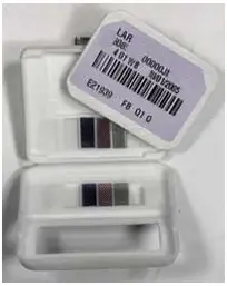
Film Badge. Source: www.nde-ed.org
Film badges are small portable devices for monitoring cumulative radiation dose due to ionizing radiation. Principle of operation is similar as for X-ray pictures. The badge consists of two parts: photographic film, and a holder. The film is contained inside a badge. The piece of photographic film that is the sensitive material and it must be removed monthly and developed. The more radiation exposure, the more blackening of the film. The blackening of the film is linear to the dose, and doses up to about 10 Gy can be measured.
See also: The Radiation Dosimeters for Response and Recovery Market Survey Report. National Urban Security Technology Laboratory. SAVER-T-MSR-4. <available from: https://www.dhs.gov/sites/default/files/publications/Radiation-Dosimeters-Response-Recovery-MSR_0616-508_0.pdf>.
Robert Reed Burn, Introduction to Nuclear Reactor Operation, 1988.
U.S. Department of Energy, Nuclear Physics and Reactor Theory. DOE Fundamentals Handbook, Volume 1 and 2. January 1993.
Paul Reuss, Neutron Physics. EDP Sciences, 2008. ISBN: 978-2759800414.
See also:
Radiation Dosimetry
We hope, this article, Active Dosimeter – Passive Dosimeter, helps you. If so, give us a like in the sidebar. Main purpose of this website is to help the public to learn some interesting and important information about radiation and dosimeters.
Dosimetry in Nuclear Power Plants. Thera are many devices (detectors) used for dosimetry. These detectors also also categorized according to their purpose. It must be noted, the following devices are not necessary dosimeters. Radiation Dosimetry
Ionizing radiation – hazard symbol
Radiation dosimetry is the measurement, calculation and assessment of the absorbed doses and assigning those doses to individuals. It is the science and practice that attempts to quantitatively relate specific measures made in a radiation field to chemical and/or biological changes that the radiation would produce in a target. Since there are two types of radiation exposure, external and internal exposure, dosimetry may be also categorized as:
External Dosimetry. External exposure is radiation that comes from outside our body and interacts with us. In this case, we analyze predominantly exposure from gamma rays and beta particles, since alpha particles, in general, constitute no external exposure hazard because the particles generally do not pass through skin. Since photons and beta interact through electromagnetic forces and neutrons interact through nuclear forces, their detection methods and dosimetry are substantially different. The source of radiation can be, for example, a piece of equipment that produces the radiation like a container with a radioactive materials, or like an x-ray machine. External dosimetry is based on measurements with a dosimeter, or inferred from measurements made by other radiological protection instruments.
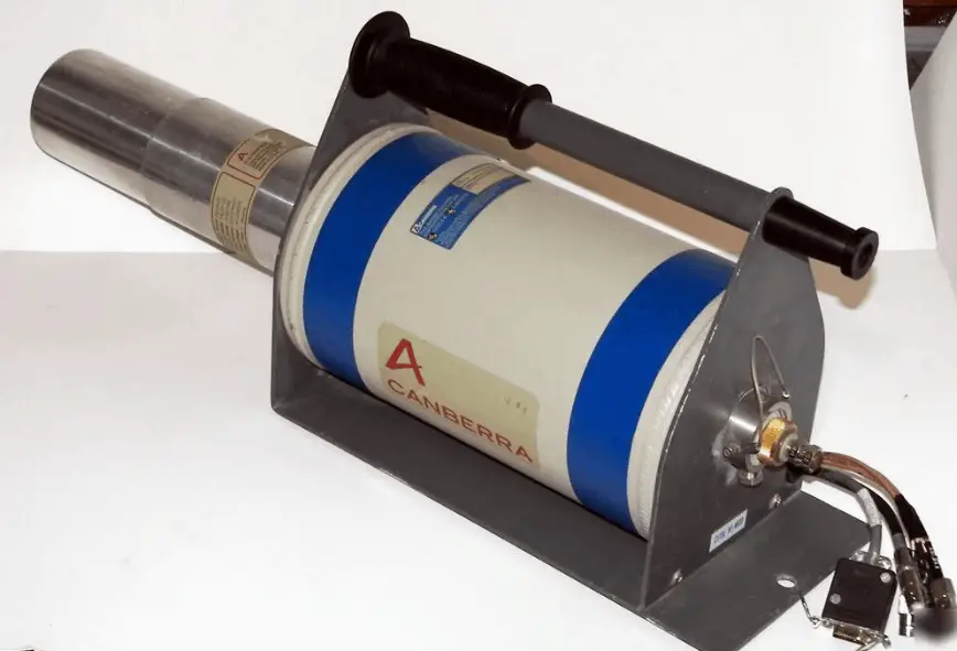
HPGe detector with LN2 cryostat, which can be used in whole-body counters. Source: canberra.com
Internal Dosimetry. If the source of radiation is inside our body, we say, it is internal exposure. The intake of radioactive material can occur through various pathways such as ingestion of radioactive contamination in food or liquids. Protection from internal exposure is more complicated. Most radionuclides will give you much more radiation dose if they can somehow enter your body, than they would if they remained outside. Internal dosimetry assessment relies on a variety of monitoring, bio-assay or radiation imaging techniques.
Detectors used in Nuclear Power Plants
Radiation dosimeters and detectors can be also categorized according to their purpose. It must be noted, the following devices are not necessary dosimeters. These devices are used for dosimetry in nuclear power plants:
Personal Dosimeters. Personal dosimetry is a key part of radiation dosimetry. Personal dosimetry is used primarily (but not exclusively) to determine doses to individuals who are exposed to radiation related to their work activities. These doses are usually measured by devices known as personal dosimeters.
Gamma Survey Meters. Portable survey meters are radiation detectors used by radiological technicians to measure ambient dose rate. These portable instruments usually have rate meters. In nuclear facilities, these portable survey meters are typically used by radiation protection technicians.
Contamination Meters. Contamination meters are instruments for surface contamination measurement. In nuclear facilities, contamination monitors are installed usually at the exit from the controlled areas. These monitors may utilize proportional counters with a large area, thin window detector similar to Hand & Shoe Monitors.
Full-Body Monitors. Full-Body Monitors, or Whole-Body Monitors, are instruments for surface contamination measurement. They are used for personnel exit monitoring, which is the term used in radiation protection for checking for external contamination (or surface contamination) of a whole body of a person leaving a radioactive contamination controlled area.
Gamma Spectrometers. A gamma ray spectrometer (GRS) is a sophisticated device for measuring the energy distribution of gamma radiation. For the measurement of gamma rays above several hundred keV, there are two detector categories of major importance, inorganic scintillators as NaI(Tl) and semiconductor detectors.
Radiation Monitoring System. At nuclear facilities, remote radiation monitoring systems (RMS) are installed to monitor radiation levels at selected plant locations. The radiation monitoring system with pre-set alarm levels (e.g. for dose, dose rate or airborne activity) provide a reliable means of real time monitoring of the radiological conditions to which a worker is exposed. If these levels are exceeded, alarms are activated and in some cases automatic protective functions initiated. Thus the system serves to:
Warn of any radiation health hazard
Give an early warning of a plant malfunction
Initiate automatic protective functions.
All data are collected in a radiation protection control room. The radiation monitoring system may gather all information on the radiological conditions at various working areas, as well as voice and visual feedback, with a minimum presence of RP technicians in radiation areas, therefore reducing dose to such personnel. Several types of radiation monitors are used in the RMS, depending on the source and strength of the radiation source.
Airborne Contamination Monitors
Area Monitors
Iodine Monitors
In-vent Gas Monitors
It is very important, that most of personal dosimeters in use today are not absolute instruments, but reference instruments. That means , they must be periodically calibrated. When a reference dosimeter is calibrated, a calibration factor can be determined. This calibration factor relates the exposure quantity to the reported dose. Validity of the calibration is demonstrated by maintaining traceability of the source used to calibrate the dosimeter. The traceability is achieved by comparison of the source with a “primary standard” at a reference calibration centre. In monitoring of individuals, the values of these operational quantities are taken as a sufficiently precise assessment of effective dose and skin dose, respectively, in particular, if their values are below the protection limits.
Characteristics of Dosimeters – Key Features
There are many types of dosimeters and detectors, and each type has limitations. Many factors influence the quality of a dosimeter’s results. Some key considerations when choosing a dosimeter include:
Type of radiation. Each type of radiation interacts with matter in a different way. This consideration is crucial. For doses from neutrons, we cannot use a simple GM counter.
Energy of radiation. A dosimeter’s response will vary depending on the energy of the radiation and the angle(s) between the source and the dosimeter’s detector.
Fading. A dosimeter’s signal can be lost or fade over time. This can be caused by external factors such as temperature, light and humidity.
Direct reading. Sometimes, it is of the highest importance, that the dosimeter can give a continuous readout of cumulative dose and current dose rate, and can warn the person wearing it when a specified dose rate or a cumulative dose is exceeded.
Minimum measurable dose. The lowest dose that can be measured with a certain specified confidence level.
Ruggedness and ease of wear. Dosimeters differ in their ability to withstand severe environmental conditions. Some heavy for a given purpose, some are smaller, lighter and more portable.
As can be seen, radiation dosimetry is very difficult, since no single dosimeter will have every one of these characteristics. Therefore, a dosimeter user must understand the environment where the instrument will be used. In most practical situations, dosimeters provide reasonable approximations to the personal dose equivalent, Hp(d), at least at the location of the dosimeter. It must be noted, the personal dose equivalent generally overestimates the effective dose. On the other hand, this procedure is valid only at low doses and under the assumption of a uniform whole-body exposure. For high personal doses approaching or exceeding the annual dose limit, or in strongly inhomogeneous radiation fields, however, this procedure might not be sufficient.
See also: The Radiation Dosimeters for Response and Recovery Market Survey Report. National Urban Security Technology Laboratory. SAVER-T-MSR-4. <available from: https://www.dhs.gov/sites/default/files/publications/Radiation-Dosimeters-Response-Recovery-MSR_0616-508_0.pdf>.
Types of Dosimeters
Film Badge Dosimeters
Film badges are small portable devices for monitoring cumulative radiation dose due to ionizing radiation. Principle of operation is similar as for X-ray pictures. The badge consists of two parts: photographic film, and a holder. The film is contained inside a badge. The piece of photographic film that is the sensitive material and it must be removed monthly and developed. The more radiation exposure, the more blackening of the film. The blackening of the film is linear to the dose, and doses up to about 10 Gy can be measured.
See also: Film Badge Dosimeter
TLD – Thermoluminescent Dosimeter
A thermoluminescent dosimeter, abbreviated as TLD, is a passive radiation dosimeter, that measures ionizing radiation exposure by measuring the intensity of visible light emitted from a sensitive crystal in the detector when the crystal is heated. The intensity of light emitted is measure by TLD reader and it is dependent upon the radiation exposure. Thermoluminescent dosimeters was invented in 1954 by Professor Farrington Daniels of the University of Wisconsin-Madison. TLD dosimeters are applicable to situations where real-time information is not needed, but precise accumulated dose monitoring records are desired for comparison to field measurements or for assessing the potential for long term health effects.
See also: TLD – Thermoluminescent Dosimeter
EPD – Electronic Personal Dosimeter
An electronic personal dosimeter is modern dosimeter, which can give a continuous readout of cumulative dose and current dose rate, and can warn the person wearing it when a specified dose rate or a cumulative dose is exceeded. EPDs are especially useful in high dose areas where residence time of the wearer is limited due to dose constraints.
The electronic personal dosimeter, EPD, is able to display a direct reading of the detected dose or dose rate in real time. Electronic dosimeters may be used as a supplemental dosimeter as well a primary dosimeter. The passive dosimeters and the electronic personal dosimeters are often used together to complement each other.
Robert Reed Burn, Introduction to Nuclear Reactor Operation, 1988.
U.S. Department of Energy, Nuclear Physics and Reactor Theory. DOE Fundamentals Handbook, Volume 1 and 2. January 1993.
Paul Reuss, Neutron Physics. EDP Sciences, 2008. ISBN: 978-2759800414.
See also:
Radiation Dosimetry
We hope, this article, Dosimetry in Nuclear Power Plants, helps you. If so, give us a like in the sidebar. Main purpose of this website is to help the public to learn some interesting and important information about radiation and dosimeters.
Characteristics of Dosimeters – Key Features. Many factors influence the quality of a dosimeter’s results. Some key considerations when choosing a dosimeter include: type of radiation, minimum measurable dose or fading. Radiation Dosimetry
EPD – Electronic Personal Dosimeters
A radiation dosimeter is a device that measures exposure to ionizing radiation. Dosimeters usually record a dose, which is the absorbed radiation energy measured in grays (Gy) or the equivalent dose measured in sieverts (Sv). A personal dosimeter is dosimeter, that is worn at the surface of the body by the person being monitored, and it records of the radiation dose received.
Characteristics of Dosimeters – Key Features
There are many types of dosimeters and detectors, and each type has limitations. Many factors influence the quality of a dosimeter’s results. Some key considerations when choosing a dosimeter include:
Type of radiation. Each type of radiation interacts with matter in a different way. This consideration is crucial. For doses from neutrons, we cannot use a simple GM counter.
Energy of radiation. A dosimeter’s response will vary depending on the energy of the radiation and the angle(s) between the source and the dosimeter’s detector.
Fading. A dosimeter’s signal can be lost or fade over time. This can be caused by external factors such as temperature, light and humidity.
Direct reading. Sometimes, it is of the highest importance, that the dosimeter can give a continuous readout of cumulative dose and current dose rate, and can warn the person wearing it when a specified dose rate or a cumulative dose is exceeded.
Minimum measurable dose. The lowest dose that can be measured with a certain specified confidence level.
Ruggedness and ease of wear. Dosimeters differ in their ability to withstand severe environmental conditions. Some heavy for a given purpose, some are smaller, lighter and more portable.
As can be seen, radiation dosimetry is very difficult, since no single dosimeter will have every one of these characteristics. Therefore, a dosimeter user must understand the environment where the instrument will be used. In most practical situations, dosimeters provide reasonable approximations to the personal dose equivalent, Hp(d), at least at the location of the dosimeter. It must be noted, the personal dose equivalent generally overestimates the effective dose. On the other hand, this procedure is valid only at low doses and under the assumption of a uniform whole-body exposure. For high personal doses approaching or exceeding the annual dose limit, or in strongly inhomogeneous radiation fields, however, this procedure might not be sufficient.
See also: The Radiation Dosimeters for Response and Recovery Market Survey Report. National Urban Security Technology Laboratory. SAVER-T-MSR-4. <available from: https://www.dhs.gov/sites/default/files/publications/Radiation-Dosimeters-Response-Recovery-MSR_0616-508_0.pdf>.
Robert Reed Burn, Introduction to Nuclear Reactor Operation, 1988.
U.S. Department of Energy, Nuclear Physics and Reactor Theory. DOE Fundamentals Handbook, Volume 1 and 2. January 1993.
Paul Reuss, Neutron Physics. EDP Sciences, 2008. ISBN: 978-2759800414.
See also:
Radiation Dosimetry
We hope, this article, Characteristics of Dosimeters – Key Features, helps you. If so, give us a like in the sidebar. Main purpose of this website is to help the public to learn some interesting and important information about radiation and dosimeters.
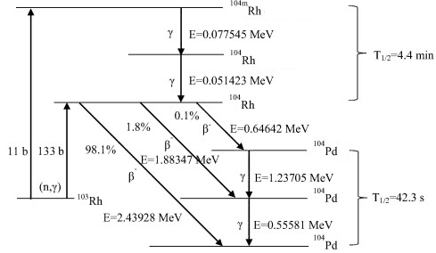
As can be seen, a neutron captured by rhodium-103 causes a rhodium-103 atom to become a radioactive rhodium-104 atom. The rhodium-104 then decays into palladium-104 plus a beta particle (electron). Rhodium-104 as Emitter – Rhodium-103 as Material
One of possible materials is rhodium as the emitter. A SPND with a rhodium emitter has a relatively high sensitivity, high burn-up rate, perturbs the local power density and has a (two-fold) delayed signal. Rhodium-based detector is the beta-current type of self-powered detector, which uses the following activation reaction to produce a current that can be measured.
1n + 103Rh → 104Rh → 104Pd + β
As can be seen, a neutron captured by rhodium-103 causes a rhodium-103 atom to become a radioactive rhodium-104 atom. The rhodium-104 then decays into palladium-104 plus a beta particle (electron). The beta particle has enough energy to pass through the insulator and reach the collector. The half-life of activated rhodium-104 is 42.3 seconds, which delays the emission of the charged particle. Rhodium based detector uses this production of beta particles (electrons) to create a current that is proportional to the number of neutrons captured by the emitter, which is also proportional to local reactor power density. A portion of the detector’s current flow is due to gamma rays. In order to compensate for this erroneous signal, a background correction is performed via background detector, which consists of the same components as the detector, except the rhodium is removed.
Rhodium-103 has a capture cross-section of 133 barns for thermal neutrons and a resonance at 1.25 eV. This reaction leads to production of 104Rh with T1/2 = 42 sec which is beta radioactive. It must be noted about 11 barns belong to reaction in which an isomer 104mRh is produced (with T1/2 = 4.4 min).
The following characteristics are typical when used in thermal power reactor (e.g. PWR).
The rhodium burnup rate is 0.39% per month in a thermal neutron flux of 1013n/cm2/sec.
Robert Reed Burn, Introduction to Nuclear Reactor Operation, 1988.
U.S. Department of Energy, Nuclear Physics and Reactor Theory. DOE Fundamentals Handbook, Volume 1 and 2. January 1993.
Paul Reuss, Neutron Physics. EDP Sciences, 2008. ISBN: 978-2759800414.
See also:
Incore Instrumentation
We hope, this article, Rhodium-104 as Emitter – Rhodium-103 as Material, helps you. If so, give us a like in the sidebar. Main purpose of this website is to help the public to learn some interesting and important information about radiation and dosimeters.
A SPND with a vanadium emitter has a relatively low sensitivity, low burn-up rate, with minimal perturbation of the local power density and has a very long delayed signal. Vanadium-51 as Material – Vanadium-based SPND
Self-Powered Neutron Detectors (SPND) are neutron detectors, that are being widely used in reactors to monitor neutron flux due to its adaptability for in-core severe environment. SPNDs may be a part of the incore neutron flux monitoring system, which provides detailed information on neutron flux distribution and thus the margins to these peak power limits. These detectors use the basic radioactive decay process of its neutron activation material to produce an output signal. As the name implies, SPNDs do not require an external voltage source to create a voltage potential in the detector. Instead, a current is produced in the detector as the result of neutron activation and subsequent beta decay of the detector itself. Because of the emission of these beta particles (electrons), the wire becomes more and more positively charged. The positive potential of the wire causes a current to flow in resistor, R. The electron current from beta decay can be measured directly with an ammeter.
There are two main advantages of the self-powered neutron detector:
Very little instrumentation is required, usually only a millivoltmeter or an ammeter
The emitter material has a much greater lifetime than boron or uranium-235 lining used in fission chambers.
On the other hand, there are also disadvantages, one is associated with the fact that currents even at full power operation are very low. Therefore, SPNDs are unable to provide with information about flux distribution at low power operation (10% and less). The main disadvantage of the self-powered neutron detector is that the emitter material decays with a characteristic half-life, which determines the response time of the detector.
The typical SPND is a coaxial cable consisting of:
Emitter. An inner electrode, which is made from a material that absorbs a neutron and undergoes radioactive decay by emitting an electron (beta decay). The emitter is usually made of rhodium and is used to produce electrons.
Insulation. The emitter is surrounded by insulation, which is usually made of aluminum oxide.
Collector. The metal walls of the detector encase these parts and serve as a collector for the. electrons that are produced.- The collector is attached to ground potential,
Self-powered neutron detectors are usually placed into the instrumentation tube of a fuel assembly, they can monitor the entire length of selected fuel assemblies to provide an extremely accurate, three-dimensional map of the neutron flux distribution. Using these data, neutron flux reconstruction can be performed also in the rest of the reactor core.
Typical materials used for the emitter are cobalt, cadmium, rhodium, and vanadium. These materials should be used because they possess relatively high melting temperatures, relatively high cross sections to thermal neutrons and are compatible with the SPND manufacturing process.
Special Reference: William H. Todt, Sr. CHARACTERISTICS OF SELF-POWERED NEUTRON DETECTORS USED IN POWER REACTORS. Imaging and Sensing Technology Corporation. New York.
Vanadium Emitter – Vanadium-based SPND
A SPND with a vanadium emitter has a relatively low sensitivity, low burn-up rate, with minimal perturbation of the local power density and has a very long delayed signal. Vanadium-based detector is the beta-current type of self-powered detector, which uses the following activation reaction to produce a current that can be measured.
1n + 51V → 52V → 52Cr + β
Vanadium-51 has a capture cross-section of 4.9 barns for thermal neutrons without resonances. This reaction leads to production of 52V with T1/2 = 3.74 min which is beta radioactive.
The following characteristics are typical when used in thermal power reactor (e.g. PWR).
The vanadium burnup rate is 0.012% per month in a thermal neutron flux of 1013n/cm2/sec.
99% of the signal has a half-life of 3.8 minutes.
1% of the signal is prompt.
The subsequent beta emission has an energy of 2.6 MeV.
Robert Reed Burn, Introduction to Nuclear Reactor Operation, 1988.
U.S. Department of Energy, Nuclear Physics and Reactor Theory. DOE Fundamentals Handbook, Volume 1 and 2. January 1993.
Paul Reuss, Neutron Physics. EDP Sciences, 2008. ISBN: 978-2759800414.
See also:
Incore Instrumentation
We hope, this article, Vanadium-51 as Material – Vanadium-based SPND, helps you. If so, give us a like in the sidebar. Main purpose of this website is to help the public to learn some interesting and important information about radiation and dosimeters.
 In radiation protection, the rem (an abbreviation for Roentgen Equivalent Man) is the non-SI unit of the equivalent dose, which is used predominantly in the USA. The rem represents the equivalent biological effect of the deposit of one hundred ergs (one rad) of gamma rays energy in a kilogram of human tissue. The rem is not derived from the unit of exposure, the roentgen. The acronym is now a misleading historical artifact, since 1 roentgen actually deposits about 0.96 rem in soft biological tissue, when all weighting factors equal unity.