Invisible radiation includes radio waves, infrared, UV, microwaves, and gamma radiation. In addition, alpha and beta radiation as well as “”cathode rays” – all of which are streams of particles – are invisible.
Source: Tour of the Electromagnetic Spectrum www.nasa.gov
In general, electromagnetic radiation may be divided into a visible and invisible portion of electromagnetic spectrum. Light is electromagnetic radiation within a certain portion of the electromagnetic spectrum. At the lower end of the visible light spectrum, electromagnetic radiation becomes invisible to humans (infrared) because its photons no longer have enough individual energy to cause a lasting molecular change (a change in conformation) in the visual molecule retinal in the human retina, which change triggers the sensation of vision.
Invisible Radiation
All electromagnetic radiation except visible light (a very narrow band) is invisible. Invisible radiation includes radio waves, infrared, UV, microwaves, and gamma radiation. In addition, alpha and beta radiation as well as “”cathode rays” – all of which are streams of particles – are invisible.
Noteworthy neither invisible radiation is completely invisible to the human eye. A related topic is that of cosmic-ray visual phenomena, in which astronauts can see flashes of light, which are presumably due to individual cosmic-ray particles interacting with their eyes. Researchers believe that these light flashes perceived specifically by astronauts in space are due to cosmic rays (high-energy charged particles from beyond the Earth’s atmosphere), though the exact mechanism is unknown.
The danger of ionizing radiation lies in the fact that the radiation is invisible and not directly detectable by human senses. People can neither see nor feel radiation, yet it deposits energy to the molecules of material. The energy is transferred in small quantities for each interaction between the radiation and a molecule and there are usually many types of interactions. Therefore, the only way you can detect and measure radiation is to use instruments (detectors of ionizing radiation).
Detection of Invisible Radiation
Detectors of ionizing radiation consist of two parts that are usually connected. The first part consists of a sensitive material, consisting of a compound that experiences changes when exposed to radiation. The other component is a device that converts these changes into measurable signals.
In their basic principles of operation, most detectors of ionizing radiation follow similar characteristics. Detectors of ionizing radiation consist of two parts that are usually connected. The first part consists of a sensitive material, consisting of a compound that experiences changes when exposed to radiation. The other component is a device that converts these changes into measurable signals. All detectors require that radiation must deposit some of its energy in sensitive material that forms part of the instrument. The radiation enters the detector, interacts with atoms of the detector material and deposits some energy to sensitive material. Each event may generate a signal, which can be a pulse, hole, light signal, ion pairs in a gas, and many others. The main task is to generate sufficient signal, amplify it and to record it.
Robert Reed Burn, Introduction to Nuclear Reactor Operation, 1988.
U.S. Department of Energy, Nuclear Physics and Reactor Theory. DOE Fundamentals Handbook, Volume 1 and 2. January 1993.
Paul Reuss, Neutron Physics. EDP Sciences, 2008. ISBN: 978-2759800414.
See also:
Visible and Invisible Radiation
We hope, this article, Invisible Radiation, helps you. If so, give us a like in the sidebar. Main purpose of this website is to help the public to learn some interesting and important information about radiation and dosimeters.
In order to detect invisible light, some detectors must be used. In their basic principles of operation, most detectors of ionizing radiation follow similar characteristics. Detection of Invisible Radiation
All electromagnetic radiation except visible light (a very narrow band) is invisible. Invisible radiation includes radio waves, infrared, UV, microwaves, and gamma radiation. In addition, alpha and beta radiation as well as “”cathode rays” – all of which are streams of particles – are invisible.
Noteworthy neither invisible radiation is completely invisible to the human eye. A related topic is that of cosmic-ray visual phenomena, in which astronauts can see flashes of light, which are presumably due to individual cosmic-ray particles interacting with their eyes. Researchers believe that these light flashes perceived specifically by astronauts in space are due to cosmic rays (high-energy charged particles from beyond the Earth’s atmosphere), though the exact mechanism is unknown.
Detection of Invisible Radiation
Detectors of ionizing radiation consist of two parts that are usually connected. The first part consists of a sensitive material, consisting of a compound that experiences changes when exposed to radiation. The other component is a device that converts these changes into measurable signals.
In their basic principles of operation, most detectors of ionizing radiation follow similar characteristics. Detectors of ionizing radiation consist of two parts that are usually connected. The first part consists of a sensitive material, consisting of a compound that experiences changes when exposed to radiation. The other component is a device that converts these changes into measurable signals. All detectors require that radiation must deposit some of its energy in sensitive material that forms part of the instrument. The radiation enters the detector, interacts with atoms of the detector material and deposits some energy to sensitive material. Each event may generate a signal, which can be a pulse, hole, light signal, ion pairs in a gas, and many others. The main task is to generate sufficient signal, amplify it and to record it.
Robert Reed Burn, Introduction to Nuclear Reactor Operation, 1988.
U.S. Department of Energy, Nuclear Physics and Reactor Theory. DOE Fundamentals Handbook, Volume 1 and 2. January 1993.
Paul Reuss, Neutron Physics. EDP Sciences, 2008. ISBN: 978-2759800414.
See also:
Visible and Invisible Radiation
We hope, this article, Detection of Invisible Radiation, helps you. If so, give us a like in the sidebar. Main purpose of this website is to help the public to learn some interesting and important information about radiation and dosimeters.
Radiation dosimetry is the measurement, calculation and assessment of the absorbed doses and assigning those doses to individuals. Radiation Dosimetry
Ionizing radiation – hazard symbol
Radiation dosimetry is the measurement, calculation and assessment of the absorbed doses and assigning those doses to individuals. It is the science and practice that attempts to quantitatively relate specific measures made in a radiation field to chemical and/or biological changes that the radiation would produce in a target.
Since there are two types of radiation exposure, external and internal exposure, dosimetry may be also categorized as:
External Dosimetry. External exposure is radiation that comes from outside our body and interacts with us. In this case, we analyze predominantly exposure from gamma rays and beta particles, since alpha particles, in general, constitute no external exposure hazard because the particles generally do not pass through skin. Since photons and beta interact through electromagnetic forces and neutrons interact through nuclear forces, their detection methods and dosimetry are substantially different. The source of radiation can be, for example, a piece of equipment that produces the radiation like a container with a radioactive materials, or like an x-ray machine. External dosimetry is based on measurements with a dosimeter, or inferred from measurements made by other radiological protection instruments.
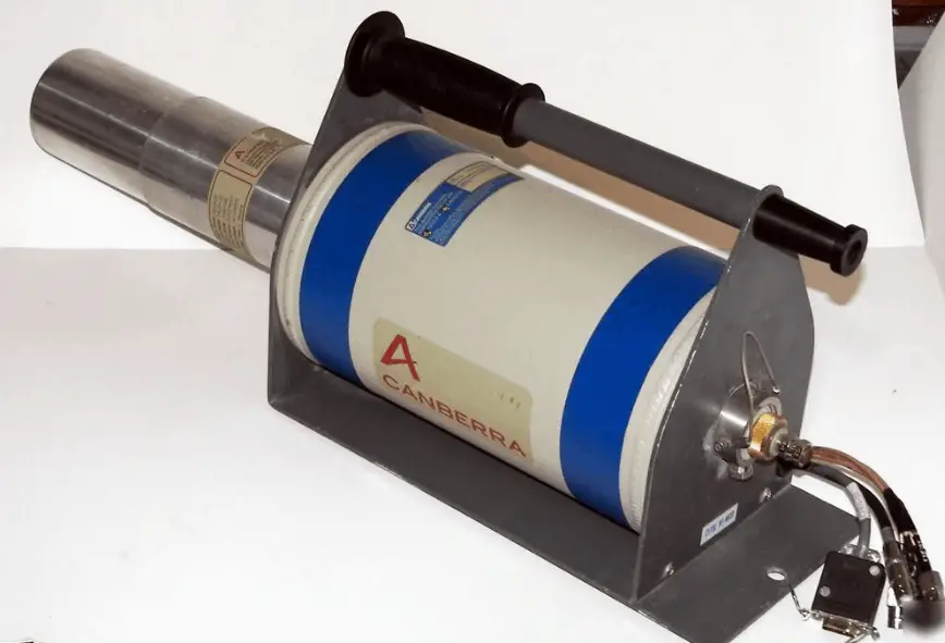
HPGe detector with LN2 cryostat, which can be used in whole-body counters. Source: canberra.com
Internal Dosimetry. If the source of radiation is inside our body, we say, it is internal exposure. The intake of radioactive material can occur through various pathways such as ingestion of radioactive contamination in food or liquids. Protection from internal exposure is more complicated. Most radionuclides will give you much more radiation dose if they can somehow enter your body, than they would if they remained outside. Internal dosimetry assessment relies on a variety of monitoring, bio-assay or radiation imaging techniques.
Personal Dosimetry
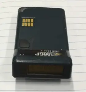
EPD – Electronic Personal Dosimeter
Personal dosimetry is a key part of radiation dosimetry. Personal dosimetry is used primarily (but not exclusively) to determine doses to individuals who are exposed to radiation related to their work activities. These doses are usually measured by devices known as dosimeters. Dosimeters usually record a dose, which is the absorbed radiation energy measured in grays (Gy) or the equivalent dose measured in sieverts (Sv). A personal dosimeter is dosimeter, that is worn at the surface of the body by the person being monitored, and it records of the radiation dose received. Personal dosimetry techniques vary and depend partly on whether the source of radiation is outside the body (external) or taken into the body (internal). Personal dosimeters are used to measure external radiation exposures. Internal exposures are typically monitored by measuring the presence of nuclear substances in the body, or by measuring nuclear substances excreted by the body.
Commercially available dosimeters range from low-cost, passive devices that store personnel dose information for later readout, to more expensive, battery operated devices that display immediate dose and dose rate information (typically an electronic personal dosimeter). Readout method, dose measurement range, size, weight, and price are important selection factors.
There are two kinds of dosimeters:
Passive Dosimeters. Commonly used passive dosimeters are the Thermo Luminescent Dosimeter (TLD) and the film badge. A passive dosimeter produces a radiation-induced signal, which is stored in the device. The dosimeter is then processed and the output is analyzed.
Active Dosimeters. To get a real time value of your exposure you can instead use an active dosimeter, typically an electronic personal dosimeter (EPD). An active dosimeter produces a radiation-induced signal and displays a direct reading of the detected dose or dose rate in real time.
The passive and the active dosimeters are often used together to complement each other. To estimate effective doses, dosimeters must be worn on a position of the body representative of its exposure, typically between the waist and the neck, on the front of the torso, facing the radioactive source. Dosimeters are usually worn on the outside of clothing, around the chest or torso to represent dose to the “whole body”. Dosimeters may also be worn on the extremities or near the eye to measure equivalent dose to these tissues.
The personal dosimeters in use today are not absolute instruments, but reference instruments. That means , they must be periodically calibrated. When a reference dosimeter is calibrated, a calibration factor can be determined. This calibration factor relates the exposure quantity to the reported dose. Validity of the calibration is demonstrated by maintaining traceability of the source used to calibrate the dosimeter. The traceability is achieved by comparison of the source with a “primary standard” at a reference calibration centre. In monitoring of individuals, the values of these operational quantities are taken as a sufficiently precise assessment of effective dose and skin dose, respectively, in particular, if their values are below the protection limits.
Example - Electronic Personal Dosimeter
EPD – Electronic Personal Dosimeter
An electronic personal dosimeter is modern dosimeter, which can give a continuous readout of cumulative dose and current dose rate, and can warn the person wearing it when a specified dose rate or a cumulative dose is exceeded. EPDs are especially useful in high dose areas where residence time of the wearer is limited due to dose constraints.
Types of EPDs
EPDs are battery powered and most use either a small Geiger-Mueller (GM) tube or a semiconductor in which ionizing radiation releases charges resulting in measurable electric current.
G-M counter. A Geiger counter consists of a Geiger-Müller tube (the sensing element which detects the radiation) and the processing electronics, which displays the result. G-M counters are mainly used for portable instrumentation due to its sensitivity, simple counting circuit, and ability to detect low-level radiation. Because of the large avalanche induced by any ionization, a Geiger counter takes a long time (about 1 ms) to recover between successive pulses. Therefore, Geiger counters are not able to measure high radiation rates due to the “dead time” of the tube.
Semiconductor Detector. Semiconductor detectors are based on ionization in a solid (e.g. silicon) and include different types of solid-state devices with two terminals called diodes. For example a silicon diode, which has a p-i-n structure in which the intrinsic (i) region is sensitive to ionizing radiation, particularly X rays and gamma rays. Under reverse bias, an electric field extends across the intrinsic or depleted region. In this case, negative voltage is applied to the p-side and positive to the second one. Holes in the p-region are attracted from the junction towards the p contact and similarly for electrons and the n contact.
Scintillation Detector. Some EPDs use a scintillating crystal such as sodium iodide (NaI) or cesium iodide (CsI) with a photodiode or photomultiplier tube to measure photons released by radiation.
Characteristics of EPDs
The electronic personal dosimeter, EPD, is able to display a direct reading of the detected dose or dose rate in real time. Electronic dosimeters may be used as a supplemental dosimeter as well a primary dosimeter. The passive dosimeters and the electronic personal dosimeters are often used together to complement each other. To estimate effective doses, dosimeters must be worn on a position of the body representative of its exposure, typically between the waist and the neck, on the front of the torso, facing the radioactive source. Dosimeters are usually worn on the outside of clothing, around the chest or torso to represent dose to the “whole body”. Dosimeters may also be worn on the extremities or near the eye to measure equivalent dose to these tissues.
The dosimeter can be reset, usually after taking a reading for record purposes, and thereby re-used multiple times. The EPDs have a top mounted display to make them easy to read when they are clipped to your breast pocket. The digital display gives both dose and dose rate information usually in mSv and mSv/h. The EPD has a dose rate alarm, and a dose alarm. These alarms are programmable. Different alarms can be set for different activities.
For example:
dose rate alarm at 100 μSv/h,
dose alarm: 100 μSv.
If an alarm set point is reached, the relevant display flashes along with a red light, and quite a piercing noise is generated. You can clear the dose rate alarm by retreating to a lower radiation field, but you cannot clear the dose alarm until you get to a EPD reader. EPDs can also give a bleep for every 1 or 10 μSv they register. This gives you an audible indication of the radiation fields. Some EPDs have wireless communication capabilities. EPDs are capable of measuring a wide radiation dose range from routine (μSv) levels to emergency levels (hundreds mSv or units of Sieverts) with high precision, and may display the exposure rate as well as accumulated exposure values. Of the dosimeter technologies, electronic personal dosimeters are generally the most expensive, largest in size, and the most versatile.
DMC 3000 – Mirion Technologies Inc.
The DMC 3000 is an electronic radiation dosimeter, EPD, that provides dose and ambient dose rate readings for deep dose equivalent Hp(10). It is one of the most used EPDs on the market. It uses a Si chipdetector with gamma sensitivity of 180 cps/R/h. This electronic personal dosimeter has the following characteristics:
Energy response (X-ray and gamma) from 15 keV to 7 Mev.
Dose measurement display range: between 1 μSv and 10 Sv.
Rate measurement display range: between 10 μSv/hr and 10 Sv/h.
The device measures 3.3 x 1.9 x 0.7 inches and has options for being clipped to a pocket, belt, or lanyard. It is powered with rechargeable or AAA batteries with a battery life of up to 2,500 hours of continuous use. Audible and visual indicators signal a low battery condition. The device has a backlit, eight-digit LCD display; two-button navigation; and visual LED, audible, and vibrating alarm indicators. Calibration is expected to last 9 months under routine use and 2 years in storage. Data is stored in nonvolatile memory. The operating range for the dosimeter is from 14°F to 122°F and up to 90 percent relative humidity. It is drop tested to 1.5 meters. The DMC 3000 has optional external modules that expand the device’s detection and communication capabilities. These include a beta module that provides Hp(0,07) for beta radiation measurement; a neutron module that provides Hp(10) neutron radiation measurement; and a telemetry module that allows transmission of data to an external station.
See also: The Radiation Dosimeters for Response and Recovery Market Survey Report. National Urban Security Technology Laboratory. SAVER-T-MSR-4. <available from: https://www.dhs.gov/sites/default/files/publications/Radiation-Dosimeters-Response-Recovery-MSR_0616-508_0.pdf>.
Medical Dosimetry
Medical dosimetry is the calculation of absorbed dose and optimization of dose delivery in medical examinations and treatments. In general, radiation exposures from medical diagnostic examinations are low (especially in diagnostic uses). Doses may be also high (only for therapeutic uses), but in each case, they must be always justified by the benefits of accurate diagnosis of possible disease conditions or by benefits of accurate treatment. These doses include contributions from medical and dental diagnostic radiology (diagnostic X-rays), clinical nuclear medicine and radiation therapy. Medical dosimetry is often performed by a professional health physicist with specialized training in that field. In order to plan the delivery of radiation therapy, the radiation produced by the sources is usually characterized with percentage depth dose curves and dose profiles measured by a medical physicist.
The medical use of ionizing radiation remains a rapidly changing field. In any case, usefulness of ionizing radiation must be balanced with its hazards. Nowadays a compromise was found and most of uses of radiation are optimized. Today it is almost unbelievable that x-rays was, at one time, used to find the right pair of shoes (i.e. shoe-fitting fluoroscopy). Measurements made in recent years indicate that the doses to the feet were in the range 0.07 – 0.14 Gy for a 20 second exposure. This practice was halted when the risks of ionizing radiation were better understood.
Environmental dosimetry is used where it is likely that the environment will generate a significant radiation dose. As was written, radiation is all around us. In, around, and above the world we live in. It is a natural energy force that surrounds us. It is a part of our natural world that has been here since the birth of our planet. All living creatures, from the beginning of time, have been, and are still being, exposed to ionizing radiation. Ionizing radiation is generated through nuclear reactions, nuclear decay, by very high temperature, or via acceleration of charged particles in electromagnetic fields.
In general, there are two broad categories of radiation sources in the environment:
Natural Background Radiation. Natural background radiation includes radiation produced by the Sun, lightnings, primordial radioisotopes or supernova explosions etc.
Man-Made Sources of Radiation. Man-made sources include medical uses of radiation, residues from nuclear tests, industrial uses of radiation etc.
An example of environment dosimetry is radon monitoring. Radon is a radioactive gas generated by the decay of uranium, which is present in varying amounts in the earth’s crust. It is important to note that radon is a noble gas, whereas all its decay products are metals. The main mechanism for the entry of radon into the atmosphere is diffusion through the soil. Certain geographic areas, due to the underlying geology, continually generate radon which permeates its way to the earth’s surface. In some cases the dose can be significant in buildings where the gas can accumulate. Locations with higher radon background are well mapped in each country. In the open air, it ranges from 1 to 100 Bq/m3, even less (0.1 Bq/m3) above the ocean. In caves or aerated mines, or ill-aerated houses, its concentration climbs to 20–2,000 Bq/m3. In the outdoor atmosphere, there is also some advection caused by wind and changes in barometric pressure. A number of specialised dosimetry techniques are used to evaluate the dose that a building’s occupants may receive.
Example - Gamma Spectroscopy
Gamma Spectroscopy
As was written, the study and analysis of gamma ray spectra for scientific and technical use is called gamma spectroscopy, and gamma ray spectrometers are the instruments which observe and collect such data. A gamma ray spectrometer (GRS) is a sophisticated device for measuring the energy distribution of gamma radiation. For the measurement of gamma rays above several hundred keV, there are two detector categories of major importance, inorganic scintillators as NaI(Tl) and semiconductor detectors. In the previous articles, we described the gamma spectroscopy using scintillation detector, which consists of a suitable scintillator crystal, a photomultiplier tube, and a circuit for measuring the height of the pulses produced by the photomultiplier. The advantages of a scintillation counter are its efficiency (large size and high density) and the high precision and counting rates that are possible. Due to the high atomic number of iodine, a large number of all interactions will result in complete absorption of gamma-ray energy, so the photo fraction will be high.
HPGe detector with LN2 cryostat Source: canberra.com
But if a perfect energy resolution is required, we have to use germanium-based detector, such as the HPGe detector. Germanium-based semiconductor detectors are most commonly used where a very good energy resolution is required, especially for gamma spectroscopy, as well as x-ray spectroscopy. In gamma spectroscopy, germanium is preferred due to its atomic number being much higher than silicon and which increases the probability of gamma ray interaction. Moreover, germanium has lower average energy necessary to create an electron-hole pair, which is 3.6 eV for silicon and 2.9 eV for germanium. This also provides the latter a better resolution in energy. The FWHM (full width at half maximum) for germanium detectors is a function of energy. For a 1.3 MeV photon, the FWHM is 2.1 keV, which is very low.
Radiation Dose Measuring and Monitoring
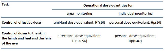
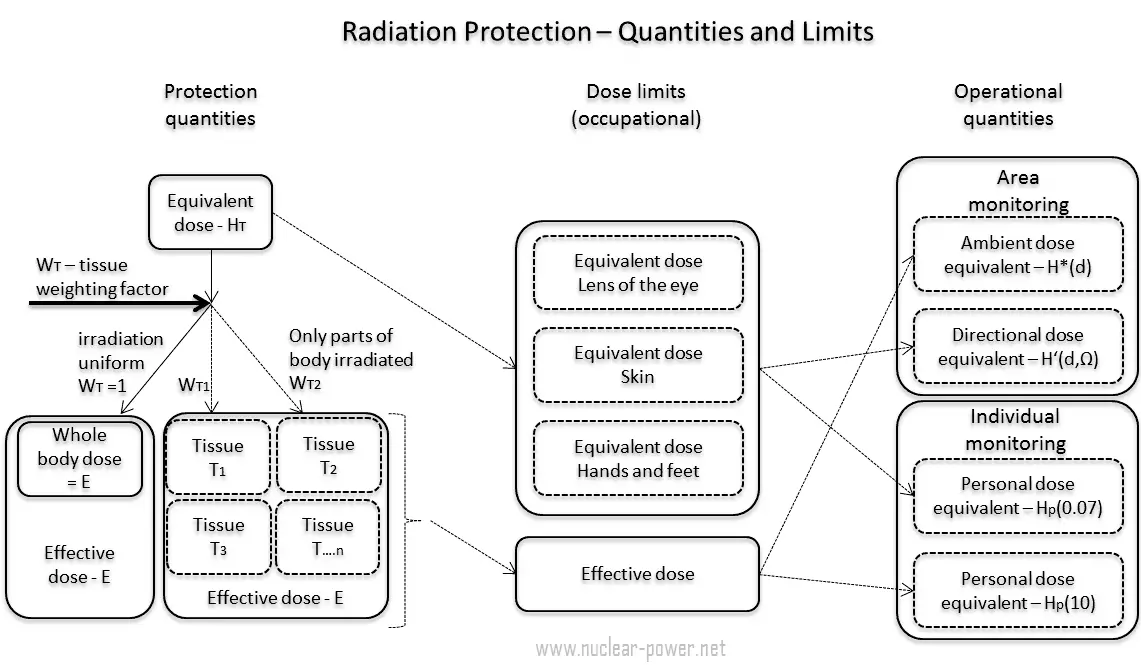
In previous chapters, we described the equivalent dose and the effective dose. But these doses are not directly measurable. For this purpose, the ICRP has introduced and defined a set of operational quantities, which can be measured and which are intended to provide a reasonable estimate for the protection quantities. These quantities aim to provide a conservative estimate for the value of the protection quantities related to an exposure avoiding both underestimation and too much overestimation.
Numerical links between these quantities is represented by conversion coefficients, which are defined for a reference person. It is very important that an internationally agreed set of conversion coefficients is available for general use in radiological protection practice for occupational exposures and exposures of the public. For the calculation of conversion coefficients for external exposure, computational phantoms are used for dose assessment in various radiation fields. For the calculation of dose coefficients from intakes of radionuclides, biokinetic models for radionuclides, reference physiological data, and computational phantoms are used.
A set of evaluated data of conversion coefficients for protection, and operational quantities for external exposure to mono-energetic photon, neutron, and electron radiation under specific irradiation conditions is published in reports (ICRP, 1996b, ICRU, 1997).
In general, the ICRP defines operational quantities for area and individual monitoring of external exposures. The operational quantities for area monitoring are:
Ambient dose equivalent, H*(10). The ambient dose equivalent is an operational quantity for area monitoring of strongly penetrating radiation.
Directional dose equivalent, H’ (d,Ω). The directional dose equivalent is an operational quantity for area monitoring of weakly penetrating radiation.
Personal dose equivalent, Hp(0.07). The Hp(0.07) dose equivalent is an operational quantity for individual monitoring for the assessment of the dose to the skin and to the hands and feet.
Personal dose equivalent, Hp(10). The Hp(10) dose equivalent is an operational quantity for individual monitoring for the assessment of effective dose.
Special Reference: ICRP, 2007. The 2007 Recommendations of the International Commission on Radiological Protection. ICRP Publication 103. Ann. ICRP 37 (2-4).
Dose limits are split into two groups, the public, and occupationally exposed workers. According to ICRP, occupational exposure refers to all exposure incurred by workers in the course of their work, with the exception of
excluded exposures and exposures from exempt activities involving radiation or exempt sources
any medical exposure
the normal local natural background radiation.
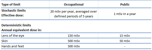
The following table summarizes dose limits for occupationally exposed workers and for the public:
Table of dose limits for occupationally exposed workers and for the public. Source of data: ICRP, 2007. The 2007 Recommendations of the International Commission on Radiological Protection. ICRP Publication 103. Ann. ICRP 37 (2-4).
According to the recommendation of the ICRP in its statement on tissue reactions of 21. April 2011, the equivalent dose limit for the lens of the eye for occupational exposure in planned exposure situations was reduced from 150 mSv/year to 20 mSv/year, averaged over defined periods of 5 years, with no annual dose in a single year exceeding 50 mSv.
Limits on effective dose are for the sum of the relevant effective doses from external exposure in the specified time period and the committed effective dose from intakes of radionuclides in the same period. For adults, the committed effective dose is computed for a 50-year period after intake, whereas for children it is computed for the period up to age 70 years. The effective whole-body dose limit of 20 mSv is an average value over five years. The real limit is 100 mSv in 5 years, with not more than 50 mSv in any one year.
Sievert – Unit of Equivalent Dose
In radiation protection, the sievert is a derived unit of equivalent dose and effective dose. The sievert represents the equivalent biological effect of the deposit of a joule of gamma rays energy in a kilogram of human tissue. Unit of sievert is of importance in radiation protection and was named after the Swedish scientist Rolf Sievert, who did a lot of the early work on radiation dosimetry in radiation therapy.
As was written, the sievert is used for radiation dose quantities such as equivalent dose and effective dose. Equivalent dose (symbol HT) is a dose quantity calculated for individual organs (index T – tissue). Equivalent dose is based on the absorbed dose to an organ, adjusted to account for the effectiveness of the type of radiation. Equivalent dose is given the symbol HT. The SI unit of HT is the sievert (Sv) or but rem (roentgen equivalent man) is still commonly used (1 Sv = 100 rem).
Examples of Doses in Sieverts
We must note that radiation is all around us. In, around, and above the world we live in. It is a natural energy force that surrounds us. It is a part of our natural world that has been here since the birth of our planet. In the following points we try to express enormous ranges of radiation exposure, which can be obtained from various sources.
0.05 µSv – Sleeping next to someone
0.09 µSv – Living within 30 miles of a nuclear power plant for a year
0.1 µSv – Eating one banana
0.3 µSv – Living within 50 miles of a coal power plant for a year
10 µSv – Average daily dose received from natural background
20 µSv – Chest X-ray
40 µSv – A 5-hour airplane flight
600 µSv – mammogram
1 000 µSv – Dose limit for individual members of the public, total effective dose per annum
3 650 µSv – Average yearly dose received from natural background
5 800 µSv – Chest CT scan
10 000 µSv – Average yearly dose received from natural background in Ramsar, Iran
20 000 µSv – single full-body CT scan
175 000 µSv – Annual dose from natural radiation on a monazite beach near Guarapari, Brazil.
5 000 000 µSv – Dose that kills a human with a 50% risk within 30 days (LD50/30), if the dose is received over a very short duration.
Robert Reed Burn, Introduction to Nuclear Reactor Operation, 1988.
U.S. Department of Energy, Nuclear Physics and Reactor Theory. DOE Fundamentals Handbook, Volume 1 and 2. January 1993.
Paul Reuss, Neutron Physics. EDP Sciences, 2008. ISBN: 978-2759800414.
See also:
Nuclear Engineering
We hope, this article, Radiation Dosimetry, helps you. If so, give us a like in the sidebar. Main purpose of this website is to help the public to learn some interesting and important information about radiation and dosimeters.
External dosimetry is based on measurements with a dosimeter, or inferred from measurements made by other radiological protection instruments. Radiation Dosimetry
Ionizing radiation – hazard symbol
Radiation dosimetry is the measurement, calculation and assessment of the absorbed doses and assigning those doses to individuals. It is the science and practice that attempts to quantitatively relate specific measures made in a radiation field to chemical and/or biological changes that the radiation would produce in a target.
External Dosimetry
External exposure is radiation that comes from outside our body and interacts with us. In this case, we analyze predominantly exposure from gamma rays and beta particles, since alpha particles, in general, constitute no external exposure hazard because the particles generally do not pass through skin. Since photons and beta interact through electromagnetic forces and neutrons interact through nuclear forces, their detection methods and dosimetry are substantially different. The source of radiation can be, for example, a piece of equipment that produces the radiation like a container with a radioactive materials, or like an x-ray machine. External dosimetry is based on measurements with a dosimeter, or inferred from measurements made by other radiological protection instruments.
Personal Dosimetry
EPD – Electronic Personal Dosimeter
Personal dosimetry is a key part of external radiation dosimetry. Personal dosimetry is used primarily (but not exclusively) to determine doses to individuals who are exposed to radiation related to their work activities. These doses are usually measured by devices known as dosimeters. Dosimeters usually record a dose, which is the absorbed radiation energy measured in grays (Gy) or the equivalent dose measured in sieverts (Sv). A personal dosimeter is dosimeter, that is worn at the surface of the body by the person being monitored, and it records of the radiation dose received. Personal dosimetry techniques vary and depend partly on whether the source of radiation is outside the body (external) or taken into the body (internal). Personal dosimeters are used to measure external radiation exposures. Internal exposures are typically monitored by measuring the presence of nuclear substances in the body, or by measuring nuclear substances excreted by the body.
Commercially available dosimeters range from low-cost, passive devices that store personnel dose information for later readout, to more expensive, battery operated devices that display immediate dose and dose rate information (typically an electronic personal dosimeter). Readout method, dose measurement range, size, weight, and price are important selection factors.
Protection against External Exposure
In radiation protection there are three ways how to protect people from identified external radiation sources:
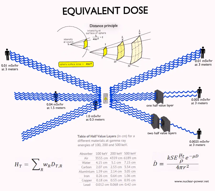
Principles of Radiation Protection – Time, Distance, Shielding
Limiting Time. The amount of radiation exposure depends directly (linearly) on the time people spend near the source of radiation. The dose can be reduced by limiting exposure time.
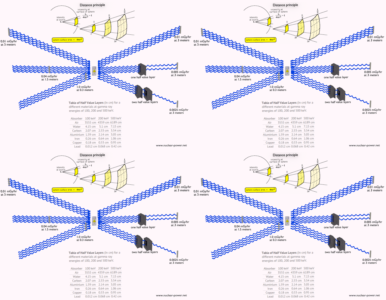
Distance. The amount of radiation exposure depends on the distance from the source of radiation. Similarly to a heat from a fire, if you are too close, the intensity of heat radiation is high and you can get burned. If you are at the right distance, you can withstand there without any problems and moreover it is comfortable. If you are too far from heat source, the insufficiency of heat can also hurt you. This analogy, in a certain sense, can be applied to radiation also from radiation sources.
Shielding. Finally, if the source is too intensive and time or distance do not provide sufficient radiation protection, the shielding must be used. Radiation shielding usually consist of barriers of lead, concrete or water. There are many many materials, which can be used for radiation shielding, but there are many many situations in radiation protection. It highly depends on the type of radiation to be shielded, its energy and many other parametres. For example, even depleted uranium can be used as a good protection from gamma radiation, but on the other hand uranium is absolutely inappropriate shielding of neutron radiation.
Robert Reed Burn, Introduction to Nuclear Reactor Operation, 1988.
U.S. Department of Energy, Nuclear Physics and Reactor Theory. DOE Fundamentals Handbook, Volume 1 and 2. January 1993.
Paul Reuss, Neutron Physics. EDP Sciences, 2008. ISBN: 978-2759800414.
See also:
Radiation Dosimetry
We hope, this article, External Dosimetry, helps you. If so, give us a like in the sidebar. Main purpose of this website is to help the public to learn some interesting and important information about radiation and dosimeters.
Internal dosimetry assessment relies on a variety of monitoring, bio-assay or radiation imaging techniques. Internal Dosimetry – Internal Exposure
Ionizing radiation – hazard symbol
Radiation dosimetry is the measurement, calculation and assessment of the absorbed doses and assigning those doses to individuals. It is the science and practice that attempts to quantitatively relate specific measures made in a radiation field to chemical and/or biological changes that the radiation would produce in a target.
Internal Dosimetry
If the source of radiation is inside our body, we say, it is internal exposure. The intake of radioactive material can occur through various pathways such as ingestion of radioactive contamination in food or liquids. Protection from internal exposure is more complicated. Most radionuclides will give you much more radiation dose if they can somehow enter your body, than they would if they remained outside. Internal dosimetry assessment relies on a variety of monitoring, bio-assay or radiation imaging techniques.
Medical dosimetry is the calculation of absorbed dose and optimization of dose delivery in medical examinations and treatments. In general, radiation exposures from medical diagnostic examinations are low (especially in diagnostic uses). Doses may be also high (only for therapeutic uses), but in each case, they must be always justified by the benefits of accurate diagnosis of possible disease conditions or by benefits of accurate treatment. These doses include contributions from medical and dental diagnostic radiology (diagnostic X-rays), clinical nuclear medicine and radiation therapy. Medical dosimetry is often performed by a professional health physicist with specialized training in that field. In order to plan the delivery of radiation therapy, the radiation produced by the sources is usually characterized with percentage depth dose curves and dose profiles measured by a medical physicist.
The medical use of ionizing radiation remains a rapidly changing field. In any case, usefulness of ionizing radiation must be balanced with its hazards. Nowadays a compromise was found and most of uses of radiation are optimized. Today it is almost unbelievable that x-rays was, at one time, used to find the right pair of shoes (i.e. shoe-fitting fluoroscopy). Measurements made in recent years indicate that the doses to the feet were in the range 0.07 – 0.14 Gy for a 20 second exposure. This practice was halted when the risks of ionizing radiation were better understood.
A whole-body counter is an instrument that measures the amounts of gamma-emitting radionuclides in the body (i.e. it is a gamma spectrometer). In nuclear facilities, these counters are used for measurement of radioactivity within the human body, that means, for internal contamination measurements. This must not be confused with a “whole body monitor” which used for personnel exit monitoring, which is the term used in radiation protection for checking for external contamination of a whole body of a person leaving a radioactive contamination controlled area. Whole-body counters are very sensitive devices and therefore they are often surrounded by large quantities of lead shielding to reduce the background radiation. A whole-body counter consists, for example, of a stand-up booth with two large-area NaI scintillation detectors. The upper detector monitors lungs, the lower detector monitors the gastrointestinal tract.
It must be noted, all people also have some radioactive isotopes inside their bodies from birth. These isotopes are especially potassium-40, carbon-14 and also the isotopes of uranium and thorium. The average annual radiation dose to a person from internal radioactive materials other than radon is about 0.3 mSv/year of which:
2 mSv/year comes from potassium-40,
12 mSv/year comes from the uranium and thorium series,
12 μSv/year comes from carbon-40.
The variation in radiation dose from one person to another is not as great, but it is detected also by a whole-body counter.
Gamma Spectroscopy
As was written, the study and analysis of gamma ray spectra for scientific and technical use is called gamma spectroscopy, and gamma ray spectrometers are the instruments which observe and collect such data. A gamma ray spectrometer (GRS) is a sophisticated device for measuring the energy distribution of gamma radiation. For the measurement of gamma rays above several hundred keV, there are two detector categories of major importance, inorganic scintillators as NaI(Tl) and semiconductor detectors. In the previous articles, we described the gamma spectroscopy using scintillation detector, which consists of a suitable scintillator crystal, a photomultiplier tube, and a circuit for measuring the height of the pulses produced by the photomultiplier. The advantages of a scintillation counter are its efficiency (large size and high density) and the high precision and counting rates that are possible. Due to the high atomic number of iodine, a large number of all interactions will result in complete absorption of gamma-ray energy, so the photo fraction will be high.
HPGe detector with LN2 cryostat Source: canberra.com
But if a perfect energy resolution is required, we have to use germanium-based detector, such as the HPGe detector. Germanium-based semiconductor detectors are most commonly used where a very good energy resolution is required, especially for gamma spectroscopy, as well as x-ray spectroscopy. In gamma spectroscopy, germanium is preferred due to its atomic number being much higher than silicon and which increases the probability of gamma ray interaction. Moreover, germanium has lower average energy necessary to create an electron-hole pair, which is 3.6 eV for silicon and 2.9 eV for germanium. This also provides the latter a better resolution in energy. The FWHM (full width at half maximum) for germanium detectors is a function of energy. For a 1.3 MeV photon, the FWHM is 2.1 keV, which is very low.
Robert Reed Burn, Introduction to Nuclear Reactor Operation, 1988.
U.S. Department of Energy, Nuclear Physics and Reactor Theory. DOE Fundamentals Handbook, Volume 1 and 2. January 1993.
Paul Reuss, Neutron Physics. EDP Sciences, 2008. ISBN: 978-2759800414.
See also:
Radiation Dosimetry
We hope, this article, Internal Dosimetry, helps you. If so, give us a like in the sidebar. Main purpose of this website is to help the public to learn some interesting and important information about radiation and dosimeters.
Personal dosimetry is a key part of radiation dosimetry. Personal dosimetry is used primarily (but not exclusively) to determine doses to individuals who are exposed to radiation related to their work activities. Radiation Dosimetry
Radiation dosimetry is the measurement, calculation and assessment of the absorbed doses and assigning those doses to individuals. It is the science and practice that attempts to quantitatively relate specific measures made in a radiation field to chemical and/or biological changes that the radiation would produce in a target.
Personal Dosimetry
EPD – Electronic Personal Dosimeter
Personal dosimetry is a key part of radiation dosimetry. Personal dosimetry is used primarily (but not exclusively) to determine doses to individuals who are exposed to radiation related to their work activities. These doses are usually measured by devices known as dosimeters. Dosimeters usually record a dose, which is the absorbed radiation energy measured in grays (Gy) or the equivalent dose measured in sieverts (Sv). A personal dosimeter is dosimeter, that is worn at the surface of the body by the person being monitored, and it records of the radiation dose received. Personal dosimetry techniques vary and depend partly on whether the source of radiation is outside the body (external) or taken into the body (internal). Personal dosimeters are used to measure external radiation exposures. Internal exposures are typically monitored by measuring the presence of nuclear substances in the body, or by measuring nuclear substances excreted by the body.
Commercially available dosimeters range from low-cost, passive devices that store personnel dose information for later readout, to more expensive, battery operated devices that display immediate dose and dose rate information (typically an electronic personal dosimeter). Readout method, dose measurement range, size, weight, and price are important selection factors.
There are two kinds of dosimeters:
Passive Dosimeters. Commonly used passive dosimeters are the Thermo Luminescent Dosimeter (TLD) and the film badge. A passive dosimeter produces a radiation-induced signal, which is stored in the device. The dosimeter is then processed and the output is analyzed.
Active Dosimeters. To get a real time value of your exposure you can instead use an active dosimeter, typically an electronic personal dosimeter (EPD). An active dosimeter produces a radiation-induced signal and displays a direct reading of the detected dose or dose rate in real time.
The passive and the active dosimeters are often used together to complement each other. To estimate effective doses, dosimeters must be worn on a position of the body representative of its exposure, typically between the waist and the neck, on the front of the torso, facing the radioactive source. Dosimeters are usually worn on the outside of clothing, around the chest or torso to represent dose to the “whole body”. Dosimeters may also be worn on the extremities or near the eye to measure equivalent dose to these tissues.
The personal dosimeters in use today are not absolute instruments, but reference instruments. That means , they must be periodically calibrated. When a reference dosimeter is calibrated, a calibration factor can be determined. This calibration factor relates the exposure quantity to the reported dose. Validity of the calibration is demonstrated by maintaining traceability of the source used to calibrate the dosimeter. The traceability is achieved by comparison of the source with a “primary standard” at a reference calibration centre. In monitoring of individuals, the values of these operational quantities are taken as a sufficiently precise assessment of effective dose and skin dose, respectively, in particular, if their values are below the protection limits.
Example – Electronic Personal Dosimeter
An electronic personal dosimeter is modern dosimeter, which can give a continuous readout of cumulative dose and current dose rate, and can warn the person wearing it when a specified dose rate or a cumulative dose is exceeded. EPDs are especially useful in high dose areas where residence time of the wearer is limited due to dose constraints.
Types of EPDs
EPDs are battery powered and most use either a small Geiger-Mueller (GM) tube or a semiconductor in which ionizing radiation releases charges resulting in measurable electric current.
G-M counter. A Geiger counter consists of a Geiger-Müller tube (the sensing element which detects the radiation) and the processing electronics, which displays the result. G-M counters are mainly used for portable instrumentation due to its sensitivity, simple counting circuit, and ability to detect low-level radiation. Because of the large avalanche induced by any ionization, a Geiger counter takes a long time (about 1 ms) to recover between successive pulses. Therefore, Geiger counters are not able to measure high radiation rates due to the “dead time” of the tube.
Semiconductor Detector. Semiconductor detectors are based on ionization in a solid (e.g. silicon) and include different types of solid-state devices with two terminals called diodes. For example a silicon diode, which has a p-i-n structure in which the intrinsic (i) region is sensitive to ionizing radiation, particularly X rays and gamma rays. Under reverse bias, an electric field extends across the intrinsic or depleted region. In this case, negative voltage is applied to the p-side and positive to the second one. Holes in the p-region are attracted from the junction towards the p contact and similarly for electrons and the n contact.
Scintillation Detector. Some EPDs use a scintillating crystal such as sodium iodide (NaI) or cesium iodide (CsI) with a photodiode or photomultiplier tube to measure photons released by radiation.
Characteristics of EPDs
The electronic personal dosimeter, EPD, is able to display a direct reading of the detected dose or dose rate in real time. Electronic dosimeters may be used as a supplemental dosimeter as well a primary dosimeter. The passive dosimeters and the electronic personal dosimeters are often used together to complement each other. To estimate effective doses, dosimeters must be worn on a position of the body representative of its exposure, typically between the waist and the neck, on the front of the torso, facing the radioactive source. Dosimeters are usually worn on the outside of clothing, around the chest or torso to represent dose to the “whole body”. Dosimeters may also be worn on the extremities or near the eye to measure equivalent dose to these tissues.
The dosimeter can be reset, usually after taking a reading for record purposes, and thereby re-used multiple times. The EPDs have a top mounted display to make them easy to read when they are clipped to your breast pocket. The digital display gives both dose and dose rate information usually in mSv and mSv/h. The EPD has a dose rate alarm, and a dose alarm. These alarms are programmable. Different alarms can be set for different activities.
For example:
dose rate alarm at 100 μSv/h,
dose alarm: 100 μSv.
If an alarm set point is reached, the relevant display flashes along with a red light, and quite a piercing noise is generated. You can clear the dose rate alarm by retreating to a lower radiation field, but you cannot clear the dose alarm until you get to a EPD reader. EPDs can also give a bleep for every 1 or 10 μSv they register. This gives you an audible indication of the radiation fields. Some EPDs have wireless communication capabilities. EPDs are capable of measuring a wide radiation dose range from routine (μSv) levels to emergency levels (hundreds mSv or units of Sieverts) with high precision, and may display the exposure rate as well as accumulated exposure values. Of the dosimeter technologies, electronic personal dosimeters are generally the most expensive, largest in size, and the most versatile.
DMC 3000 – Mirion Technologies Inc.
The DMC 3000 is an electronic radiation dosimeter, EPD, that provides dose and ambient dose rate readings for deep dose equivalent Hp(10). It is one of the most used EPDs on the market. It uses a Si chipdetector with gamma sensitivity of 180 cps/R/h. This electronic personal dosimeter has the following characteristics:
Energy response (X-ray and gamma) from 15 keV to 7 Mev.
Dose measurement display range: between 1 μSv and 10 Sv.
Rate measurement display range: between 10 μSv/hr and 10 Sv/h.
The device measures 3.3 x 1.9 x 0.7 inches and has options for being clipped to a pocket, belt, or lanyard. It is powered with rechargeable or AAA batteries with a battery life of up to 2,500 hours of continuous use. Audible and visual indicators signal a low battery condition. The device has a backlit, eight-digit LCD display; two-button navigation; and visual LED, audible, and vibrating alarm indicators. Calibration is expected to last 9 months under routine use and 2 years in storage. Data is stored in nonvolatile memory. The operating range for the dosimeter is from 14°F to 122°F and up to 90 percent relative humidity. It is drop tested to 1.5 meters. The DMC 3000 has optional external modules that expand the device’s detection and communication capabilities. These include a beta module that provides Hp(0,07) for beta radiation measurement; a neutron module that provides Hp(10) neutron radiation measurement; and a telemetry module that allows transmission of data to an external station.
See also: The Radiation Dosimeters for Response and Recovery Market Survey Report. National Urban Security Technology Laboratory. SAVER-T-MSR-4. <available from: https://www.dhs.gov/sites/default/files/publications/Radiation-Dosimeters-Response-Recovery-MSR_0616-508_0.pdf>.
Radiation Dose Measuring and Monitoring
In previous chapters, we described the equivalent dose and the effective dose. But these doses are not directly measurable. For this purpose, the ICRP has introduced and defined a set of operational quantities, which can be measured and which are intended to provide a reasonable estimate for the protection quantities. These quantities aim to provide a conservative estimate for the value of the protection quantities related to an exposure avoiding both underestimation and too much overestimation.
Numerical links between these quantities is represented by conversion coefficients, which are defined for a reference person. It is very important that an internationally agreed set of conversion coefficients is available for general use in radiological protection practice for occupational exposures and exposures of the public. For the calculation of conversion coefficients for external exposure, computational phantoms are used for dose assessment in various radiation fields. For the calculation of dose coefficients from intakes of radionuclides, biokinetic models for radionuclides, reference physiological data, and computational phantoms are used.
A set of evaluated data of conversion coefficients for protection, and operational quantities for external exposure to mono-energetic photon, neutron, and electron radiation under specific irradiation conditions is published in reports (ICRP, 1996b, ICRU, 1997).
In general, the ICRP defines operational quantities for area and individual monitoring of external exposures. The operational quantities for area monitoring are:
Ambient dose equivalent, H*(10). The ambient dose equivalent is an operational quantity for area monitoring of strongly penetrating radiation.
Directional dose equivalent, H’ (d,Ω). The directional dose equivalent is an operational quantity for area monitoring of weakly penetrating radiation.
Personal dose equivalent, Hp(0.07). The Hp(0.07) dose equivalent is an operational quantity for individual monitoring for the assessment of the dose to the skin and to the hands and feet.
Personal dose equivalent, Hp(10). The Hp(10) dose equivalent is an operational quantity for individual monitoring for the assessment of effective dose.
Special Reference: ICRP, 2007. The 2007 Recommendations of the International Commission on Radiological Protection. ICRP Publication 103. Ann. ICRP 37 (2-4).
Dose limits are split into two groups, the public, and occupationally exposed workers. According to ICRP, occupational exposure refers to all exposure incurred by workers in the course of their work, with the exception of
excluded exposures and exposures from exempt activities involving radiation or exempt sources
any medical exposure
the normal local natural background radiation.
The following table summarizes dose limits for occupationally exposed workers and for the public:
Table of dose limits for occupationally exposed workers and for the public. Source of data: ICRP, 2007. The 2007 Recommendations of the International Commission on Radiological Protection. ICRP Publication 103. Ann. ICRP 37 (2-4).
According to the recommendation of the ICRP in its statement on tissue reactions of 21. April 2011, the equivalent dose limit for the lens of the eye for occupational exposure in planned exposure situations was reduced from 150 mSv/year to 20 mSv/year, averaged over defined periods of 5 years, with no annual dose in a single year exceeding 50 mSv.
Limits on effective dose are for the sum of the relevant effective doses from external exposure in the specified time period and the committed effective dose from intakes of radionuclides in the same period. For adults, the committed effective dose is computed for a 50-year period after intake, whereas for children it is computed for the period up to age 70 years. The effective whole-body dose limit of 20 mSv is an average value over five years. The real limit is 100 mSv in 5 years, with not more than 50 mSv in any one year.
Robert Reed Burn, Introduction to Nuclear Reactor Operation, 1988.
U.S. Department of Energy, Nuclear Physics and Reactor Theory. DOE Fundamentals Handbook, Volume 1 and 2. January 1993.
Paul Reuss, Neutron Physics. EDP Sciences, 2008. ISBN: 978-2759800414.
See also:
Radiation Dosimetry
We hope, this article, Personal Dosimetry, helps you. If so, give us a like in the sidebar. Main purpose of this website is to help the public to learn some interesting and important information about radiation and dosimeters.
Medical dosimetry is the calculation of absorbed dose and optimization of dose delivery in medical examinations and treatments. Medical Dosimetry
Ionizing radiation – hazard symbol
Radiation dosimetry is the measurement, calculation and assessment of the absorbed doses and assigning those doses to individuals. It is the science and practice that attempts to quantitatively relate specific measures made in a radiation field to chemical and/or biological changes that the radiation would produce in a target.
Medical Dosimetry
Medical dosimetry is the calculation of absorbed dose and optimization of dose delivery in medical examinations and treatments. In general, radiation exposures from medical diagnostic examinations are low (especially in diagnostic uses). Doses may be also high (only for therapeutic uses), but in each case, they must be always justified by the benefits of accurate diagnosis of possible disease conditions or by benefits of accurate treatment. These doses include contributions from medical and dental diagnostic radiology (diagnostic X-rays), clinical nuclear medicine and radiation therapy. Medical dosimetry is often performed by a professional health physicist with specialized training in that field. In order to plan the delivery of radiation therapy, the radiation produced by the sources is usually characterized with percentage depth dose curves and dose profiles measured by a medical physicist.
The medical use of ionizing radiation remains a rapidly changing field. In any case, usefulness of ionizing radiation must be balanced with its hazards. Nowadays a compromise was found and most of uses of radiation are optimized. Today it is almost unbelievable that x-rays was, at one time, used to find the right pair of shoes (i.e. shoe-fitting fluoroscopy). Measurements made in recent years indicate that the doses to the feet were in the range 0.07 – 0.14 Gy for a 20 second exposure. This practice was halted when the risks of ionizing radiation were better understood.
In the following points we try to express enormous ranges of radiation exposure as well as a few doses from medical sources.
1 µSv – Eating one banana
1 µSv – Extremity (hand, foot, etc.) X-ray
5 µSv – Dental X-ray
10 µSv – Average daily dose received from natural background
40 µSv – A 5-hour airplane flight
100 µSv – Chest X-ray
600 µSv – mammogram
1 000 µSv – Dose limit for individual members of the public, total effective dose per annum
3 650 µSv – Average yearly dose received from natural background
5 800 µSv – Chest CT scan
10 000 µSv – Average yearly dose received from natural background in Ramsar, Iran
20 000 µSv – single full-body CT scan
80 000 µSv – The annual local dose to localized spots at the bifurcations of segmental bronchi in the lungs caused by smoking cigarettes (1.5 packs/day).
175 000 µSv – Annual dose from natural radiation on a monazite beach near Guarapari, Brazil.
5 000 000 µSv – Dose that kills a human with a 50% risk within 30 days (LD50/30), if the dose is received over a very short duration.
As can be seen, low-level doses are common for everyday life.
Robert Reed Burn, Introduction to Nuclear Reactor Operation, 1988.
U.S. Department of Energy, Nuclear Physics and Reactor Theory. DOE Fundamentals Handbook, Volume 1 and 2. January 1993.
Paul Reuss, Neutron Physics. EDP Sciences, 2008. ISBN: 978-2759800414.
See also:
Radiation Dosimetry
We hope, this article, Medical Dosimetry, helps you. If so, give us a like in the sidebar. Main purpose of this website is to help the public to learn some interesting and important information about radiation and dosimeters.
Environmental dosimetry is used where it is likely that the environment will generate a significant radiation dose. Environmental Dosimetry
Ionizing radiation – hazard symbol
Radiation dosimetry is the measurement, calculation and assessment of the absorbed doses and assigning those doses to individuals. It is the science and practice that attempts to quantitatively relate specific measures made in a radiation field to chemical and/or biological changes that the radiation would produce in a target.
Environmental Dosimetry
Environmental dosimetry is used where it is likely that the environment will generate a significant radiation dose. As was written, radiation is all around us. In, around, and above the world we live in. It is a natural energy force that surrounds us. It is a part of our natural world that has been here since the birth of our planet. All living creatures, from the beginning of time, have been, and are still being, exposed to ionizing radiation. Ionizing radiation is generated through nuclear reactions, nuclear decay, by very high temperature, or via acceleration of charged particles in electromagnetic fields.
In general, there are two broad categories of radiation sources in the environment:
Natural Background Radiation. Natural background radiation includes radiation produced by the Sun, lightnings, primordial radioisotopes or supernova explosions etc.
Man-Made Sources of Radiation. Man-made sources include medical uses of radiation, residues from nuclear tests, industrial uses of radiation etc.
An example of environment dosimetry is radon monitoring. Radon is a radioactive gas generated by the decay of uranium, which is present in varying amounts in the earth’s crust. It is important to note that radon is a noble gas, whereas all its decay products are metals. The main mechanism for the entry of radon into the atmosphere is diffusion through the soil. Certain geographic areas, due to the underlying geology, continually generate radon which permeates its way to the earth’s surface. In some cases the dose can be significant in buildings where the gas can accumulate. Locations with higher radon background are well mapped in each country. In the open air, it ranges from 1 to 100 Bq/m3, even less (0.1 Bq/m3) above the ocean. In caves or aerated mines, or ill-aerated houses, its concentration climbs to 20–2,000 Bq/m3. In the outdoor atmosphere, there is also some advection caused by wind and changes in barometric pressure. A number of specialised dosimetry techniques are used to evaluate the dose that a building’s occupants may receive.
Gamma Spectroscopy
As was written, the study and analysis of gamma ray spectra for scientific and technical use is called gamma spectroscopy, and gamma ray spectrometers are the instruments which observe and collect such data. A gamma ray spectrometer (GRS) is a sophisticated device for measuring the energy distribution of gamma radiation. For the measurement of gamma rays above several hundred keV, there are two detector categories of major importance, inorganic scintillators as NaI(Tl) and semiconductor detectors. In the previous articles, we described the gamma spectroscopy using scintillation detector, which consists of a suitable scintillator crystal, a photomultiplier tube, and a circuit for measuring the height of the pulses produced by the photomultiplier. The advantages of a scintillation counter are its efficiency (large size and high density) and the high precision and counting rates that are possible. Due to the high atomic number of iodine, a large number of all interactions will result in complete absorption of gamma-ray energy, so the photo fraction will be high.
HPGe detector with LN2 cryostat Source: canberra.com
But if a perfect energy resolution is required, we have to use germanium-based detector, such as the HPGe detector. Germanium-based semiconductor detectors are most commonly used where a very good energy resolution is required, especially for gamma spectroscopy, as well as x-ray spectroscopy. In gamma spectroscopy, germanium is preferred due to its atomic number being much higher than silicon and which increases the probability of gamma ray interaction. Moreover, germanium has lower average energy necessary to create an electron-hole pair, which is 3.6 eV for silicon and 2.9 eV for germanium. This also provides the latter a better resolution in energy. The FWHM (full width at half maximum) for germanium detectors is a function of energy. For a 1.3 MeV photon, the FWHM is 2.1 keV, which is very low.
Robert Reed Burn, Introduction to Nuclear Reactor Operation, 1988.
U.S. Department of Energy, Nuclear Physics and Reactor Theory. DOE Fundamentals Handbook, Volume 1 and 2. January 1993.
Paul Reuss, Neutron Physics. EDP Sciences, 2008. ISBN: 978-2759800414.
See also:
Radiation Dosimetry
We hope, this article, Environmental Dosimetry, helps you. If so, give us a like in the sidebar. Main purpose of this website is to help the public to learn some interesting and important information about radiation and dosimeters.
A radiation dosimeter is a device that measures exposure to ionizing radiation. Dosimeters usually record a dose, which is the absorbed radiation energy measured in grays (Gy) or the equivalent dose measured in sieverts (Sv). Radiation Dosimetry
A radiation dosimeter is a device that measures exposure to ionizing radiation. Dosimeters usually record a dose, which is the absorbed radiation energy measured in grays (Gy) or the equivalent dose measured in sieverts (Sv). A personal dosimeter is dosimeter, that is worn at the surface of the body by the person being monitored, and it records of the radiation dose received.
EPD – Electronic Personal Dosimeters
Commercially available dosimeters range from low-cost, passive devices that store personnel dose information for later readout, to more expensive, battery operated devices that display immediate dose and dose rate information (typically an electronic personal dosimeter). Readout method, dose measurement range, size, weight, and price are important selection factors.
There are two kinds of dosimeters:
Passive Dosimeters. Commonly used passive dosimeters are the Thermo Luminescent Dosimeter (TLD) and the film badge. A passive dosimeter produces a radiation-induced signal, which is stored in the device. The dosimeter is then processed and the output is analyzed.
Active Dosimeters. To get a real time value of your exposure you can instead use an active dosimeter, typically an electronic personal dosimeter (EPD). An active dosimeter produces a radiation-induced signal and displays a direct reading of the detected dose or dose rate in real time.
The passive and the active dosimeters are often used together to complement each other. To estimate effective doses, dosimeters must be worn on a position of the body representative of its exposure, typically between the waist and the neck, on the front of the torso, facing the radioactive source. Dosimeters are usually worn on the outside of clothing, around the chest or torso to represent dose to the “whole body”. Dosimeters may also be worn on the extremities or near the eye to measure equivalent dose to these tissues.
Radiation dosimeters and detectors can be also categorized according to their purpose. It must be noted, the following devices are not necessary dosimeters. These devices are used for dosimetry in nuclear power plants:
Personal Dosimeters. Personal dosimetry is a key part of radiation dosimetry. Personal dosimetry is used primarily (but not exclusively) to determine doses to individuals who are exposed to radiation related to their work activities. These doses are usually measured by devices known as personal dosimeters.
Gamma Survey Meters. Portable survey meters are radiation detectors used by radiological technicians to measure ambient dose rate. These portable instruments usually have rate meters. In nuclear facilities, these portable survey meters are typically used by radiation protection technicians.
Contamination Meters. Contamination meters are instruments for surface contamination measurement. In nuclear facilities, contamination monitors are installed usually at the exit from the controlled areas. These monitors may utilize proportional counters with a large area, thin window detector similar to Hand & Shoe Monitors.
Full-Body Monitors. Full-Body Monitors, or Whole-Body Monitors, are instruments for surface contamination measurement. They are used for personnel exit monitoring, which is the term used in radiation protection for checking for external contamination (or surface contamination) of a whole body of a person leaving a radioactive contamination controlled area.
Gamma Spectrometers. A gamma ray spectrometer (GRS) is a sophisticated device for measuring the energy distribution of gamma radiation. For the measurement of gamma rays above several hundred keV, there are two detector categories of major importance, inorganic scintillators as NaI(Tl) and semiconductor detectors.
It is very important, that most of personal dosimeters in use today are not absolute instruments, but reference instruments. That means , they must be periodically calibrated. When a reference dosimeter is calibrated, a calibration factor can be determined. This calibration factor relates the exposure quantity to the reported dose. Validity of the calibration is demonstrated by maintaining traceability of the source used to calibrate the dosimeter. The traceability is achieved by comparison of the source with a “primary standard” at a reference calibration centre. In monitoring of individuals, the values of these operational quantities are taken as a sufficiently precise assessment of effective dose and skin dose, respectively, in particular, if their values are below the protection limits.
Characteristics of Dosimeters – Key Features
There are many types of dosimeters and detectors, and each type has limitations. Many factors influence the quality of a dosimeter’s results. Some key considerations when choosing a dosimeter include:
Type of radiation. Each type of radiation interacts with matter in a different way. This consideration is crucial. For doses from neutrons, we cannot use a simple GM counter.
Energy of radiation. A dosimeter’s response will vary depending on the energy of the radiation and the angle(s) between the source and the dosimeter’s detector.
Fading. A dosimeter’s signal can be lost or fade over time. This can be caused by external factors such as temperature, light and humidity.
Direct reading. Sometimes, it is of the highest importance, that the dosimeter can give a continuous readout of cumulative dose and current dose rate, and can warn the person wearing it when a specified dose rate or a cumulative dose is exceeded.
Minimum measurable dose. The lowest dose that can be measured with a certain specified confidence level.
Ruggedness and ease of wear. Dosimeters differ in their ability to withstand severe environmental conditions. Some heavy for a given purpose, some are smaller, lighter and more portable.
As can be seen, radiation dosimetry is very difficult, since no single dosimeter will have every one of these characteristics. Therefore, a dosimeter user must understand the environment where the instrument will be used. In most practical situations, dosimeters provide reasonable approximations to the personal dose equivalent, Hp(d), at least at the location of the dosimeter. It must be noted, the personal dose equivalent generally overestimates the effective dose. On the other hand, this procedure is valid only at low doses and under the assumption of a uniform whole-body exposure. For high personal doses approaching or exceeding the annual dose limit, or in strongly inhomogeneous radiation fields, however, this procedure might not be sufficient.
See also: The Radiation Dosimeters for Response and Recovery Market Survey Report. National Urban Security Technology Laboratory. SAVER-T-MSR-4. <available from: https://www.dhs.gov/sites/default/files/publications/Radiation-Dosimeters-Response-Recovery-MSR_0616-508_0.pdf>.
Types of Dosimeters
Film Badge Dosimeters
Film badges are small portable devices for monitoring cumulative radiation dose due to ionizing radiation. Principle of operation is similar as for X-ray pictures. The badge consists of two parts: photographic film, and a holder. The film is contained inside a badge. The piece of photographic film that is the sensitive material and it must be removed monthly and developed. The more radiation exposure, the more blackening of the film. The blackening of the film is linear to the dose, and doses up to about 10 Gy can be measured.
See also: Film Badge Dosimeter
TLD – Thermoluminescent Dosimeter
A thermoluminescent dosimeter, abbreviated as TLD, is a passive radiation dosimeter, that measures ionizing radiation exposure by measuring the intensity of visible light emitted from a sensitive crystal in the detector when the crystal is heated. The intensity of light emitted is measure by TLD reader and it is dependent upon the radiation exposure. Thermoluminescent dosimeters was invented in 1954 by Professor Farrington Daniels of the University of Wisconsin-Madison. TLD dosimeters are applicable to situations where real-time information is not needed, but precise accumulated dose monitoring records are desired for comparison to field measurements or for assessing the potential for long term health effects.
See also: TLD – Thermoluminescent Dosimeter
EPD – Electronic Personal Dosimeter
An electronic personal dosimeter is modern dosimeter, which can give a continuous readout of cumulative dose and current dose rate, and can warn the person wearing it when a specified dose rate or a cumulative dose is exceeded. EPDs are especially useful in high dose areas where residence time of the wearer is limited due to dose constraints.
The electronic personal dosimeter, EPD, is able to display a direct reading of the detected dose or dose rate in real time. Electronic dosimeters may be used as a supplemental dosimeter as well a primary dosimeter. The passive dosimeters and the electronic personal dosimeters are often used together to complement each other.
See also: EPD – Electronic Personal Dosimeter
MOSFET Dosimeter
MOSFET dosimeter is a small portable device for monitoring and direct reading of radiation dose rate. Since it is based on the MOSFET transistor, the metal-oxide-semiconductor field-effect transistor (MOSFET), the principle of operation is similar as for semiconductor detectors. MOSFET dosimeters are now used as clinical dosimeters for radiotherapy radiation beams. Their main advantage is their physical size, which is less than 4 mm2. In radiation therapy dosimetry, MOSFET dosimeters often replace TLD dosimeters, since they offer immediate read out.
See also: MOSFET Dosimeter
Self-reading Dosimeter
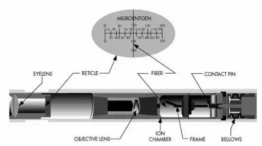
The self indicating pocket dosimeter consists of an ionization chamber, with a volume of approximately two milliliters, which is sensitive to a desired radiation, a quartz fiber electrometer to measure the charge, and a microscope to read the fiber image off a scale. Source: www.nde-ed.org
Self-reading dosimeters are field-readable devices worn on the body to measure accumulated dose. These are unpowered devices that do not contain a battery. Devices in this group include:
Quartz fiber dosimeter. A quartz fiber dosimeter, sometimes called a self indicating pocket dosimeter (SIPD), is a pen-like device that measures the cumulative dose of ionizing radiation received by the device, usually over one work period.
Self-developing photochemical cards. Self-developing photochemical card is a credit card sized, instant color developing emergency dosimeter. It is designed for monitoring exposure in a radiological incident for medical treatment triage and to minimize worry and panic.
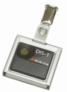
Direct-ion storage dosimeter, DIS, is an electronic dosimeter, from which the dose information for both Hp(10) and Hp(0.07) can be obtained instantly at the workplace by using an electronic reader unit. The DIS dosimeter is based on the combination of an ion chamber and a non-volatile electronic charge storage element. DIS dosimeter use an analog memory cell inside a small, gas-filled, ionization chamber. Incident radiation causes ionizations in the chamber wall and in the gas, and the charge is stored for subsequent readout. The DIS dosimeter is read at the user’s site through connection to an electronic reader unit.
Robert Reed Burn, Introduction to Nuclear Reactor Operation, 1988.
U.S. Department of Energy, Nuclear Physics and Reactor Theory. DOE Fundamentals Handbook, Volume 1 and 2. January 1993.
Paul Reuss, Neutron Physics. EDP Sciences, 2008. ISBN: 978-2759800414.
See also:
Radiation Dosimetry
We hope, this article, Radiation Dosimeter, helps you. If so, give us a like in the sidebar. Main purpose of this website is to help the public to learn some interesting and important information about radiation and dosimeters.
millisievert – microsievert. Equivalent doses measured in industry and medicine often have usually lower doses than one sievert, and the following multiples are often used: 1 mSv (millisievert) = 1E-3 Sv and 1 µSv (microsievert) = 1E-6 Sv
In radiation protection, the sievert is a derived unit of equivalent dose and effective dose. The sievert represents the equivalent biological effect of the deposit of a joule of gamma rays energy in a kilogram of human tissue. Unit of sievert is of importance in radiation protection and was named after the Swedish scientist Rolf Sievert, who did a lot of the early work on dosimetry in radiation therapy.
As was written, the sievert is used for radiation dose quantities such as equivalent dose and effective dose. Equivalent dose (symbol HT) is a dose quantity calculated for individual organs (index T – tissue). Equivalent dose is based on the absorbed dose to an organ, adjusted to account for the effectiveness of the type of radiation. Equivalent dose is given the symbol HT. The SI unit of HT is the sievert (Sv) or but rem (roentgen equivalent man) is still commonly used (1 Sv = 100 rem).
One sievert is a large amount of equivalent dose. A person who has absorbed a whole body dose of 1 Sv has absorbed one joule of energy in each kg of body tissue (in case of gamma rays).
Equivalent doses measured in industry and medicine often have usually lower doses than one sievert, and the following multiples are often used:
1 mSv (millisievert) = 1E-3 Sv
1 µSv (microsievert) = 1E-6 Sv
Conversions from the SI units to other units are as follows:
1 Sv = 100 rem
1 mSv = 100 mrem
Examples of Doses in millisieverts
We must note that radiation is all around us. In, around, and above the world we live in. It is a natural energy force that surrounds us. It is a part of our natural world that has been here since the birth of our planet. In the following points we try to express enormous ranges of radiation exposure, which can be obtained from various sources.
0.00005 mSv – Sleeping next to someone
0.00009 mSv – Living within 30 miles of a nuclear power plant for a year
0.0001 mSv – Eating one banana
0.0003 mSv – Living within 50 miles of a coal power plant for a year
0.01 mSv – Average daily dose received from natural background
0.02 mSv – Chest X-ray
0.04 mSv – A 5-hour airplane flight
0.60 mSv – mammogram
1 mSv – Dose limit for individual members of the public, total effective dose per annum
3.65 mSv – Average yearly dose received from natural background
5.8 mSv – Chest CT scan
10 mSv – Average yearly dose received from natural background in Ramsar, Iran
20 mSv – single full-body CT scan
175 mSv – Annual dose from natural radiation on a monazite beach near Guarapari, Brazil.
5 000 mSv – Dose that kills a human with a 50% risk within 30 days (LD50/30), if the dose is received over a very short duration.
Robert Reed Burn, Introduction to Nuclear Reactor Operation, 1988.
U.S. Department of Energy, Nuclear Physics and Reactor Theory. DOE Fundamentals Handbook, Volume 1 and 2. January 1993.
Paul Reuss, Neutron Physics. EDP Sciences, 2008. ISBN: 978-2759800414.
See also:
Sievert
We hope, this article, millisievert – microsievert – units of equivalent dose, helps you. If so, give us a like in the sidebar. Main purpose of this website is to help the public to learn some interesting and important information about radiation and dosimeters.
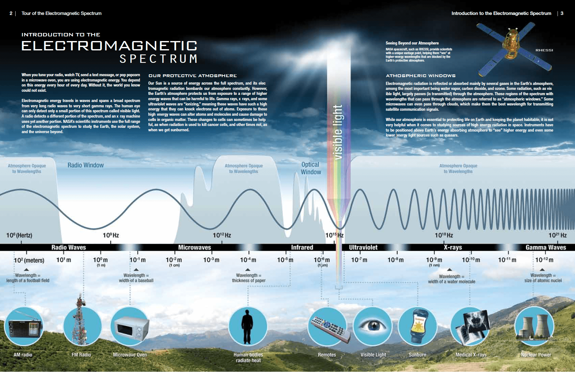
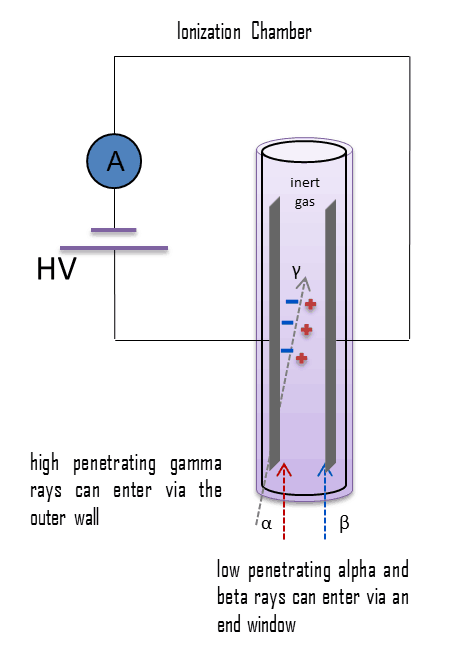 Detectors of ionizing radiation consist of two parts that are usually connected. The first part consists of a sensitive material, consisting of a compound that experiences changes when exposed to radiation. The other component is a device that converts these changes into measurable signals.