The radiation exposure rate is the rate at which an exposure is received. It is a measure of exposure intensity (or strength). It is measured in mR/sec or R/hr. Radiation Dosimetry
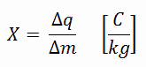
Radiation exposure is a measure of the ionization of air due to ionizing radiation from high-energy photons (i.e. X-rays and gamma rays). Radiation exposure is defined as the sum of electrical charges (∆q) on all the ions of one sign produced in air when all the electrons, liberated by photons in a volume of air whose mass is ∆m, are completely stopped in air.
Radiation exposure is given the symbol X. The SI unit of radiation exposure is the coulomb per kilogram (C/kg), but in practice, the roentgen is used.
Radiation Exposure Rate
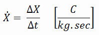
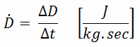
The radiation exposure rate is the rate at which an exposure is received. It is a measure of exposure intensity (or strength). The exposure rate is therefore defined as:
In conventional units, it is measured in mR/sec or R/hr. Since the amount of radiation exposure depends directly (linearly) on the time people spend near the source of radiation, the exposure is equal to the strength of the radiation field (exposure rate) multiplied by the length of time spent in that field. It must be emphasized that this concept applies only to X-rays or γ-rays in air at a point outside a body. Radiation exposure is not applicable to describe neutrons, charged particles or all interactions taking place within a body.
Robert Reed Burn, Introduction to Nuclear Reactor Operation, 1988.
U.S. Department of Energy, Nuclear Physics and Reactor Theory. DOE Fundamentals Handbook, Volume 1 and 2. January 1993.
Paul Reuss, Neutron Physics. EDP Sciences, 2008. ISBN: 978-2759800414.
See also:
Exposure
We hope, this article, Radiation Exposure Rate, helps you. If so, give us a like in the sidebar. Main purpose of this website is to help the public to learn some interesting and important information about radiation and dosimeters.
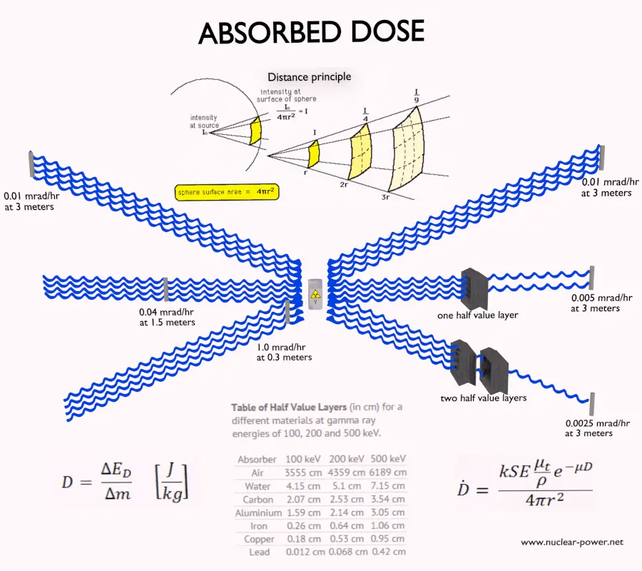
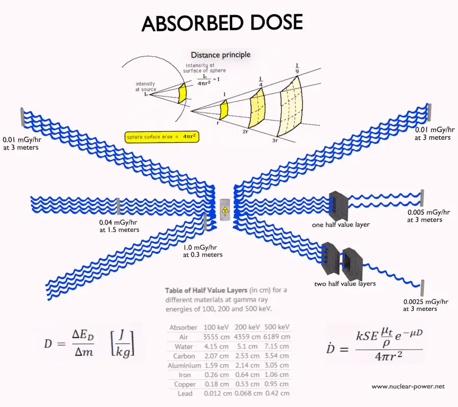
The absorbed dose rate is the rate at which an absorbed dose is received. It is a measure of radiation dose intensity (or strength). Absorbed Dose Rate
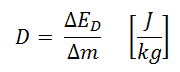
Absorbed dose is defined as the amount of energy deposited by ionizing radiation in a substance. Absorbed dose is given the symbol D. The absorbed dose is usually measured in a unit called the gray (Gy), which is derived from the SI system. The non-SI unit rad is sometimes also used, predominantly in the USA.
Units of absorbed dose:
Gray. A dose of one gray is equivalent to a unit of energy (joule) deposited in a kilogram of a substance.
RAD. A dose of one rad is equivalent to the deposition of one hundred ergs of energy in one gram of any material.
Absorbed Dose Rate
The absorbed dose rate is the rate at which an absorbed dose is received. It is a measure of radiation dose intensity (or strength). The absorbed dose rate is therefore defined as:
In conventional units, it is measured in mrad/sec, rad/hr, mGy/sec or Gy/hr. Since the amount of radiation exposure depends directly (linearly) on the time people spend near the source of radiation, the absorbed dose is equal to the strength of the radiation field (dose rate) multiplied by the length of time spent in that field. The example above indicates a person could expect to receive a dose of 25 millirems by staying in a 50 millirems/hour field for thirty minutes.
Examples of Absorbed Doses in grays
We must note that radiation is all around us. In, around, and above the world we live in. It is a natural energy force that surrounds us. It is a part of our natural world that has been here since the birth of our planet. In the following points we try to express enormous ranges of radiation exposure, which can be obtained from various sources.
0.05 µGy – Sleeping next to someone
0.09 µGy – Living within 30 miles of a nuclear power plant for a year
0.1 µGy – Eating one banana
0.3 µGy – Living within 50 miles of a coal power plant for a year
10 µGy – Average daily dose received from natural background
20 µGy – Chest X-ray
40 µGy – A 5-hour airplane flight
600 µGy – mammogram
1 000 µGy – Dose limit for individual members of the public, total effective dose per annum
3 650 µGy – Average yearly dose received from natural background
5 800 µGy – Chest CT scan
10 000 µGy – Average yearly dose received from natural background in Ramsar, Iran
20 000 µGy – single full-body CT scan
175 000 µGy – Annual dose from natural radiation on a monazite beach near Guarapari, Brazil.
5 000 000 µGy – Dose that kills a human with a 50% risk within 30 days (LD50/30), if the dose is received over a very short duration.
As can be seen, low-level doses are common for everyday life. The previous examples can help illustrate relative magnitudes. From biological consequences point of view, it is very important to distinguish between doses received over short and extended periods. An “acute dose” is one that occurs over a short and finite period of time, while a “chronic dose” is a dose that continues for an extended period of time so that it is better described by a dose rate. High doses tend to kill cells, while low doses tend to damage or change them. Low doses spread out over long periods of time don’t cause an immediate problem to any body organ. The effects of low doses of radiation occur at the level of the cell, and the results may not be observed for many years.
Calculation of Shielded Dose Rate
Assume the point isotropic source which contains 1.0 Ci of 137Cs, which has a half-life of 30.2 years. Note that the relationship between half-life and the amount of a radionuclide required to give an activity of one curie is shown below. This amount of material can be calculated using λ, which is the decay constant of certain nuclide:
About 94.6 percent decays by beta emission to a metastable nuclear isomer of barium: barium-137m. The main photon peak of Ba-137m is 662 keV. For this calculation, assume that all decays go through this channel.
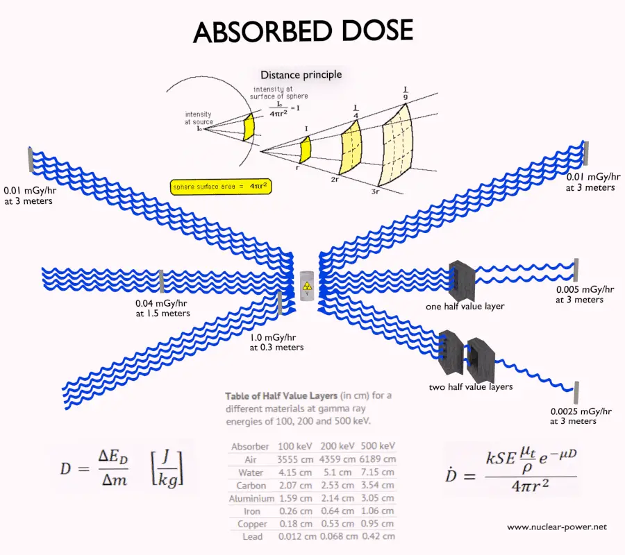
Determine the primary photon dose rate, in gray per hour (Gy.h-1), at the outer surface of a 5 cm thick lead shield. Primary photon dose rate neglects all secondary particles. Assume that the effective distance of the source from the dose point is 10 cm. We shall also assume that the dose point is soft tissue and it can reasonably be simulated by water and we use the mass energy absorption coefficient for water.
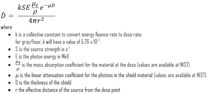
The primary photon dose rate is attenuated exponentially, and the dose rate from primary photons, taking account of the shield, is given by:
As can be seen, we do not account for the buildup of secondary radiation. If secondary particles are produced or if the primary radiation changes its energy or direction, then the effective attenuation will be much less. This assumption generally underestimates the true dose rate, especially for thick shields and when the dose point is close to the shield surface, but this assumption simplifies all calculations. For this case the true dose rate (with the buildup of secondary radiation) will be more than two times higher.
To calculate the absorbed dose rate, we have to use in the formula:
k = 5.76 x 10⁻⁷
S = 3.7 x 10¹⁰ s-1
E = 0.662 MeV
μt/ρ = 0.0326 cm2/g (values are available at NIST)
μ = 1.289 cm-1 (values are available at NIST)
D = 5 cm
r = 10 cm
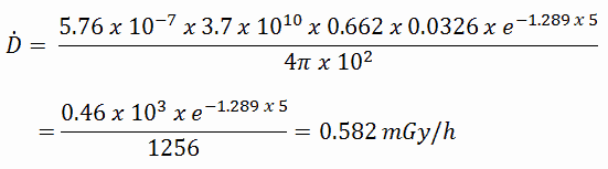
Result:
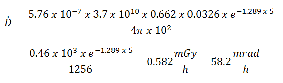
The resulting absorbed dose rate in grays per hour is then:
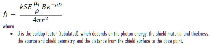
If we want to account for the buildup of secondary radiation, then we have to include the buildup factor. The extended formula for the dose rate is then:
Robert Reed Burn, Introduction to Nuclear Reactor Operation, 1988.
U.S. Department of Energy, Nuclear Physics and Reactor Theory. DOE Fundamentals Handbook, Volume 1 and 2. January 1993.
Paul Reuss, Neutron Physics. EDP Sciences, 2008. ISBN: 978-2759800414.
See also:
Absorbed Dose
We hope, this article, Absorbed Dose Rate, helps you. If so, give us a like in the sidebar. Main purpose of this website is to help the public to learn some interesting and important information about radiation and dosimeters.
The rad (an abbreviation for Radiation Absorbed Dose) is the non-SI unit of the absorbed dose. A dose of one rad is equivalent to the deposition of one hundred ergs of energy in one gram of any material. Radiation Dosimetry
Absorbed dose is defined as the amount of energy deposited by ionizing radiation in a substance. Absorbed dose is given the symbol D. The rad (an abbreviation for Radiation Absorbed Dose) is the non-SI unit of the absorbed dose. The absorbed dose is also measured in a unit called the gray (Gy), which is derived from the SI system. The non-SI unit rad is used predominantly in the USA.
Units of Absorbed Dose:
Gray. A dose of one gray is equivalent to a unit of energy (joule) deposited in a kilogram of a substance.
RAD. A dose of one rad is equivalent to the deposition of one hundred ergs of energy in one gram of any material.
RAD – Unit of Absorbed Dose
A dose of one rad is equivalent to the deposition of one hundred ergs of energy in one gram of any material. Note that, the erg is a unit of energy and work equal to 10−7 joules. A related unit, the roentgen, is used to quantify the radiation exposure. The F-factor can be used to convert between rads and roentgens.
One rad is significantly lower dose than one gray, which is a large amount of absorbed dose. A person who has absorbed a whole body dose of 100 rad has absorbed one joule of energy in each kg of body tissue (i.e. 1 Gy). Absorbed doses measured in industry (except nuclear medicine) often have comparable doses to one rad and the following multiples are often used:
1 mrad (millirad) = 1E-3 rad
1 krad (kilorad) = 1E3 rad
Conversions from the SI units to other units are as follows:
1 Gy = 100 rad
1 mGy = 100 mrad
The gray and rad are physical units. They describe the physical effect of the incident radiation (i.e., the amount of energy deposited per kg), but it tells us nothing about the biological consequences of such energy deposition in living tissue.
Examples of Absorbed Doses in rads
We must note that radiation is all around us. In, around, and above the world we live in. It is a natural energy force that surrounds us. It is a part of our natural world that has been here since the birth of our planet. In the following points we try to express enormous ranges of radiation exposure, which can be obtained from various sources.
0.005 mrad – Sleeping next to someone
0.009 mrad – Living within 30 miles of a nuclear power plant for a year
0.01 mrad – Eating one banana
0.03 mrad – Living within 50 miles of a coal power plant for a year
1 mrad – Average daily dose received from natural background
2 mrad – Chest X-ray
4 mrad – A 5-hour airplane flight
60 mrad – mammogram
100 mrad – Dose limit for individual members of the public, total effective dose per annum
365 mrad – Average yearly dose received from natural background
580 mrad – Chest CT scan
1 000 mrad – Average yearly dose received from natural background in Ramsar, Iran
2 000 mrad – single full-body CT scan
17 500 mrad – Annual dose from natural radiation on a monazite beach near Guarapari, Brazil.
500 000 mrad – Dose required to kill a human with a 50% risk within 30 days (LD50/30), if the dose is received over a very short duration.
As can be seen, low-level doses are common for everyday life. The previous examples can help illustrate relative magnitudes. From biological consequences point of view, it is very important to distinguish between doses received over short and extended periods. An “acute dose” is one that occurs over a short and finite period of time, while a “chronic dose” is a dose that continues for an extended period of time so that it is better described by a dose rate. High doses tend to kill cells, while low doses tend to damage or change them. Low doses spread out over long periods of time don’t cause an immediate problem to any body organ. The effects of low doses of radiation occur at the level of the cell, and the results may not be observed for many years.
Calculation of Shielded Dose Rate in rads
Assume the point isotropic source which contains 1.0 Ci of 137Cs, which has a half-life of 30.2 years. Note that the relationship between half-life and the amount of a radionuclide required to give an activity of one curie is shown below. This amount of material can be calculated using λ, which is the decay constant of certain nuclide:
About 94.6 percent decays by beta emission to a metastable nuclear isomer of barium: barium-137m. The main photon peak of Ba-137m is 662 keV. For this calculation, assume that all decays go through this channel.
Calculate the primary photon dose rate, in rads per hour (rad.h-1), at the outer surface of a 5 cm thick lead shield. Primary photon dose rate neglects all secondary particles. Assume that the effective distance of the source from the dose point is 10 cm. We shall also assume that the dose point is soft tissue and it can reasonably be simulated by water and we use the mass energy absorption coefficient for water.
The primary photon dose rate is attenuated exponentially, and the dose rate from primary photons, taking account of the shield, is given by:
As can be seen, we do not account for the buildup of secondary radiation. If secondary particles are produced or if the primary radiation changes its energy or direction, then the effective attenuation will be much less. This assumption generally underestimates the true dose rate, especially for thick shields and when the dose point is close to the shield surface, but this assumption simplifies all calculations. For this case the true dose rate (with the buildup of secondary radiation) will be more than two times higher.
To calculate the absorbed dose rate, we have to use in the formula:
k = 5.76 x 10⁻⁷
S = 3.7 x 10¹⁰ s-1
E = 0.662 MeV
μt/ρ = 0.0326 cm2/g (values are available at NIST)
μ = 1.289 cm-1 (values are available at NIST)
D = 5 cm
r = 10 cm
Result:
The resulting absorbed dose rate in grays per hour is then:
If we want to account for the buildup of secondary radiation, then we have to include the buildup factor. The extended formula for the dose rate is then:
Robert Reed Burn, Introduction to Nuclear Reactor Operation, 1988.
U.S. Department of Energy, Nuclear Physics and Reactor Theory. DOE Fundamentals Handbook, Volume 1 and 2. January 1993.
Paul Reuss, Neutron Physics. EDP Sciences, 2008. ISBN: 978-2759800414.
See also:
Absorbed Dose
We hope, this article, RAD – Radiation Absorbed Dose, helps you. If so, give us a like in the sidebar. Main purpose of this website is to help the public to learn some interesting and important information about radiation and dosimeters.
rad – unit – Examples and Calculation. Calculate the primary photon dose rate, in rads per hour (rad.h-1), at the outer surface of a 5 cm thick lead shield. Radiation Dosimetry
RAD – Unit of Absorbed Dose
A dose of one rad is equivalent to the deposition of one hundred ergs of energy in one gram of any material. Note that, the erg is a unit of energy and work equal to 10−7 joules. A related unit, the roentgen, is used to quantify the radiation exposure. The F-factor can be used to convert between rads and roentgens.
Examples of Absorbed Doses in rads
We must note that radiation is all around us. In, around, and above the world we live in. It is a natural energy force that surrounds us. It is a part of our natural world that has been here since the birth of our planet. In the following points we try to express enormous ranges of radiation exposure, which can be obtained from various sources.
0.005 mrad – Sleeping next to someone
0.009 mrad – Living within 30 miles of a nuclear power plant for a year
0.01 mrad – Eating one banana
0.03 mrad – Living within 50 miles of a coal power plant for a year
1 mrad – Average daily dose received from natural background
2 mrad – Chest X-ray
4 mrad – A 5-hour airplane flight
60 mrad – mammogram
100 mrad – Dose limit for individual members of the public, total effective dose per annum
365 mrad – Average yearly dose received from natural background
580 mrad – Chest CT scan
1 000 mrad – Average yearly dose received from natural background in Ramsar, Iran
2 000 mrad – single full-body CT scan
17 500 mrad – Annual dose from natural radiation on a monazite beach near Guarapari, Brazil.
500 000 mrad – Dose required to kill a human with a 50% risk within 30 days (LD50/30), if the dose is received over a very short duration.
As can be seen, low-level doses are common for everyday life. The previous examples can help illustrate relative magnitudes. From biological consequences point of view, it is very important to distinguish between doses received over short and extended periods. An “acute dose” is one that occurs over a short and finite period of time, while a “chronic dose” is a dose that continues for an extended period of time so that it is better described by a dose rate. High doses tend to kill cells, while low doses tend to damage or change them. Low doses spread out over long periods of time don’t cause an immediate problem to any body organ. The effects of low doses of radiation occur at the level of the cell, and the results may not be observed for many years.
Calculation of Shielded Dose Rate in rads
Assume the point isotropic source which contains 1.0 Ci of 137Cs, which has a half-life of 30.2 years. Note that the relationship between half-life and the amount of a radionuclide required to give an activity of one curie is shown below. This amount of material can be calculated using λ, which is the decay constant of certain nuclide:
About 94.6 percent decays by beta emission to a metastable nuclear isomer of barium: barium-137m. The main photon peak of Ba-137m is 662 keV. For this calculation, assume that all decays go through this channel.
Calculate the primary photon dose rate, in rads per hour (rad.h-1), at the outer surface of a 5 cm thick lead shield. Primary photon dose rate neglects all secondary particles. Assume that the effective distance of the source from the dose point is 10 cm. We shall also assume that the dose point is soft tissue and it can reasonably be simulated by water and we use the mass energy absorption coefficient for water.
The primary photon dose rate is attenuated exponentially, and the dose rate from primary photons, taking account of the shield, is given by:
As can be seen, we do not account for the buildup of secondary radiation. If secondary particles are produced or if the primary radiation changes its energy or direction, then the effective attenuation will be much less. This assumption generally underestimates the true dose rate, especially for thick shields and when the dose point is close to the shield surface, but this assumption simplifies all calculations. For this case the true dose rate (with the buildup of secondary radiation) will be more than two times higher.
To calculate the absorbed dose rate, we have to use in the formula:
k = 5.76 x 10⁻⁷
S = 3.7 x 10¹⁰ s-1
E = 0.662 MeV
μt/ρ = 0.0326 cm2/g (values are available at NIST)
μ = 1.289 cm-1 (values are available at NIST)
D = 5 cm
r = 10 cm
Result:
The resulting absorbed dose rate in grays per hour is then:
If we want to account for the buildup of secondary radiation, then we have to include the buildup factor. The extended formula for the dose rate is then:
Robert Reed Burn, Introduction to Nuclear Reactor Operation, 1988.
U.S. Department of Energy, Nuclear Physics and Reactor Theory. DOE Fundamentals Handbook, Volume 1 and 2. January 1993.
Paul Reuss, Neutron Physics. EDP Sciences, 2008. ISBN: 978-2759800414.
See also:
Absorbed Dose
We hope, this article, rad – unit – Examples and Calculation, helps you. If so, give us a like in the sidebar. Main purpose of this website is to help the public to learn some interesting and important information about radiation and dosimeters.
gray – unit – Examples and Calculation. Calculate the primary photon dose rate, in grays per hour (Gy.h-1), at the outer surface of a 5 cm thick lead shield. Radiation Dosimetry
Gray – Unit of Absorbed Dose
A dose of one gray is equivalent to a unit of energy (joule) deposited in a kilogram of a substance. This unit was named in honour of Louis Harold Gray, who was one of the great pioneers in radiation biology. One gray is a large amount of absorbed dose. A person who has absorbed a whole body dose of 1 Gy has absorbed one joule of energy in each kg of body tissue.
Examples of Absorbed Doses in grays
We must note that radiation is all around us. In, around, and above the world we live in. It is a natural energy force that surrounds us. It is a part of our natural world that has been here since the birth of our planet. In the following points we try to express enormous ranges of radiation exposure, which can be obtained from various sources.
0.05 µGy – Sleeping next to someone
0.09 µGy – Living within 30 miles of a nuclear power plant for a year
0.1 µGy – Eating one banana
0.3 µGy – Living within 50 miles of a coal power plant for a year
10 µGy – Average daily dose received from natural background
20 µGy – Chest X-ray
40 µGy – A 5-hour airplane flight
600 µGy – mammogram
1 000 µGy – Dose limit for individual members of the public, total effective dose per annum
3 650 µGy – Average yearly dose received from natural background
5 800 µGy – Chest CT scan
10 000 µGy – Average yearly dose received from natural background in Ramsar, Iran
20 000 µGy – single full-body CT scan
175 000 µGy – Annual dose from natural radiation on a monazite beach near Guarapari, Brazil.
5 000 000 µGy – Dose that kills a human with a 50% risk within 30 days (LD50/30), if the dose is received over a very short duration.
As can be seen, low-level doses are common for everyday life. The previous examples can help illustrate relative magnitudes. From biological consequences point of view, it is very important to distinguish between doses received over short and extended periods. An “acute dose” is one that occurs over a short and finite period of time, while a “chronic dose” is a dose that continues for an extended period of time so that it is better described by a dose rate. High doses tend to kill cells, while low doses tend to damage or change them. Low doses spread out over long periods of time don’t cause an immediate problem to any body organ. The effects of low doses of radiation occur at the level of the cell, and the results may not be observed for many years.
Calculation of Shielded Dose Rate in grays
Assume the point isotropic source which contains 1.0 Ci of 137Cs, which has a half-life of 30.2 years. Note that the relationship between half-life and the amount of a radionuclide required to give an activity of one curie is shown below. This amount of material can be calculated using λ, which is the decay constant of certain nuclide:
About 94.6 percent decays by beta emission to a metastable nuclear isomer of barium: barium-137m. The main photon peak of Ba-137m is 662 keV. For this calculation, assume that all decays go through this channel.
Calculate the primary photon dose rate, in gray per hour (Gy.h-1), at the outer surface of a 5 cm thick lead shield. Primary photon dose rate neglects all secondary particles. Assume that the effective distance of the source from the dose point is 10 cm. We shall also assume that the dose point is soft tissue and it can reasonably be simulated by water and we use the mass energy absorption coefficient for water.
The primary photon dose rate is attenuated exponentially, and the dose rate from primary photons, taking account of the shield, is given by:
As can be seen, we do not account for the buildup of secondary radiation. If secondary particles are produced or if the primary radiation changes its energy or direction, then the effective attenuation will be much less. This assumption generally underestimates the true dose rate, especially for thick shields and when the dose point is close to the shield surface, but this assumption simplifies all calculations. For this case the true dose rate (with the buildup of secondary radiation) will be more than two times higher.
To calculate the absorbed dose rate, we have to use in the formula:
k = 5.76 x 10⁻⁷
S = 3.7 x 10¹⁰ s-1
E = 0.662 MeV
μt/ρ = 0.0326 cm2/g (values are available at NIST)
μ = 1.289 cm-1 (values are available at NIST)
D = 5 cm
r = 10 cm
Result:
The resulting absorbed dose rate in grays per hour is then:
If we want to account for the buildup of secondary radiation, then we have to include the buildup factor. The extended formula for the dose rate is then:
Robert Reed Burn, Introduction to Nuclear Reactor Operation, 1988.
U.S. Department of Energy, Nuclear Physics and Reactor Theory. DOE Fundamentals Handbook, Volume 1 and 2. January 1993.
Paul Reuss, Neutron Physics. EDP Sciences, 2008. ISBN: 978-2759800414.
See also:
Absorbed Dose
We hope, this article, gray – unit – Examples and Calculation, helps you. If so, give us a like in the sidebar. Main purpose of this website is to help the public to learn some interesting and important information about radiation and dosimeters.
Gray. A dose of one gray is equivalent to a unit of energy (joule) deposited in a kilogram of a substance. RAD. A dose of one rad is equivalent to the deposition of one hundred ergs of energy in one gram of any material. Radiation Dosimetry
Absorbed dose is defined as the amount of energy deposited by ionizing radiation in a substance. Absorbed dose is given the symbol D. The rad (an abbreviation for Radiation Absorbed Dose) is the non-SI unit of the absorbed dose. The absorbed dose is also measured in a unit called the gray (Gy), which is derived from the SI system. The non-SI unit rad is used predominantly in the USA.
Units of Absorbed Dose:
Gray. A dose of one gray is equivalent to a unit of energy (joule) deposited in a kilogram of a substance.
RAD. A dose of one rad is equivalent to the deposition of one hundred ergs of energy in one gram of any material.
RAD – Unit of Absorbed Dose
A dose of one rad is equivalent to the deposition of one hundred ergs of energy in one gram of any material. Note that, the erg is a unit of energy and work equal to 10−7 joules. A related unit, the roentgen, is used to quantify the radiation exposure. The F-factor can be used to convert between rads and roentgens.
One rad is significantly lower dose than one gray, which is a large amount of absorbed dose. A person who has absorbed a whole body dose of 100 rad has absorbed one joule of energy in each kg of body tissue (i.e. 1 Gy). Absorbed doses measured in industry (except nuclear medicine) often have comparable doses to one rad and the following multiples are often used:
1 mrad (millirad) = 1E-3 rad
1 krad (kilorad) = 1E3 rad
Conversions from the SI units to other units are as follows:
1 Gy = 100 rad
1 mGy = 100 mrad
The gray and rad are physical units. They describe the physical effect of the incident radiation (i.e., the amount of energy deposited per kg), but it tells us nothing about the biological consequences of such energy deposition in living tissue.
Gray – Unit of Absorbed Dose
A dose of one gray is equivalent to a unit of energy (joule) deposited in a kilogram of a substance. This unit was named in honour of Louis Harold Gray, who was one of the great pioneers in radiation biology. One gray is a large amount of absorbed dose. A person who has absorbed a whole body dose of 1 Gy has absorbed one joule of energy in each kg of body tissue.
Absorbed doses measured in industry (except nuclear medicine) often have usually lower doses than one gray, and the following multiples are often used:
Robert Reed Burn, Introduction to Nuclear Reactor Operation, 1988.
U.S. Department of Energy, Nuclear Physics and Reactor Theory. DOE Fundamentals Handbook, Volume 1 and 2. January 1993.
Paul Reuss, Neutron Physics. EDP Sciences, 2008. ISBN: 978-2759800414.
See also:
Absorbed Dose
We hope, this article, RAD – Gray – Conversion – Units, helps you. If so, give us a like in the sidebar. Main purpose of this website is to help the public to learn some interesting and important information about radiation and dosimeters.
Kerma is a measure of kinetic energy transferred from radiation to matter. It is an acronym for “kinetic energy released per unit mass”. Kerma is given the symbol K.
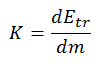
Kerma is a measure of kinetic energy transferred from radiation to matter. It is an acronym for “kinetic energy released per unit mass”. Kerma is given the symbol K and it is measured by the SI unit, the gray. This unit was named in honour of Louis Harold Gray, who was one of the great pioneers in radiation biology. Kerma is defined by the formula:
Kerma is related to, but not the same as absorbed dose. Absorbed dose is defined as the amount of energy deposited by ionizing radiation in a substance. Kerma is defined as the sum of the initial kinetic energies of all the charged particles liberated by uncharged ionizing radiation in a substance. At low energies, kerma approximately equals absorbed dose, since most of initial kinetic energies of all the charged particles deposit their energy in the sample. At higher energies kerma is larger than absorbed dose because some highly energetic secondary electrons and X-rays escape the region of interest before depositing their energy. The escaping energy is counted in kerma, but not in absorbed dose. Note that there are three key interaction mechanisms of gamma rays with matter.
Robert Reed Burn, Introduction to Nuclear Reactor Operation, 1988.
U.S. Department of Energy, Nuclear Physics and Reactor Theory. DOE Fundamentals Handbook, Volume 1 and 2. January 1993.
Paul Reuss, Neutron Physics. EDP Sciences, 2008. ISBN: 978-2759800414.
See also:
Radiation Protection
We hope, this article, Kerma – Radiation, helps you. If so, give us a like in the sidebar. Main purpose of this website is to help the public to learn some interesting and important information about radiation and dosimeters.
Interaction of X-rays with Matter. Although a large number of possible interactions are known, there are three key interaction mechanisms with matter. Radiation Dosimetry
X-rays, also known as X-radiation, refers to electromagnetic radiation (no rest mass, no charge) of high energies. X-rays are high-energy photons with short wavelengths and thus very high frequency. The radiation frequency is key parameter of all photons, because it determines the energy of a photon. Photons are categorized according to the energies from low-energy radio waves and infrared radiation, through visible light, to high-energy X-rays and gamma rays.
Most X-rays have a wavelength ranging from 0.01 to 10 nanometers (3×10¹⁶ Hz to 3×10¹⁹ Hz), corresponding to energies in the range 100 eV to 100 keV. X-ray wavelengths are shorter than those of UV rays and typically longer than those of gamma rays. The distinction between X-rays and gamma rays is not so simple and has changed in recent decades. According to the currently valid definition, X-rays are emitted by electrons outside the nucleus, while gamma rays are emitted by the nucleus.
Since the X-rays (especially hard X-rays) are in substance high-energy photons, they are very penetrating matter and are thus biologically hazardous. X-rays can travel thousands of feet in air and can easily pass through the human body.
Interaction of X-rays with Matter
Although a large number of possible interactions are known, there are three key interaction mechanisms with matter. The strength of these interactions depends on the energy of the X-rays and the elemental composition of the material, but not much on chemical properties, since the X-ray photon energy is much higher than chemical binding energies. The photoelectric absorbtion dominates at low-energies of X-rays, while Compton scattering dominates at higher energies.
Photoelectric absorption
Compton scattering
Rayleigh scattering
Photoelectric Absorption of X-rays
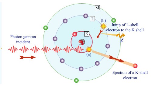
Gamma absorption by an atom. Source: laradioactivite.com/
In the photoelectric effect, a photon undergoes an interaction with an electron which is bound in an atom. In this interaction the incident photon completely disappears and an energetic photoelectron is ejected by the atom from one of its bound shells. The kinetic energy of the ejected photoelectron (Ee) is equal to the incident photon energy (hν) minus the binding energy of the photoelectron in its original shell (Eb).
Ee=hν-Eb
Therefore photoelectrons are only emitted by the photoelectric effect if photon reaches or exceeds a threshold energy – the binding energy of the electron – the work function of the material. For very high X-rays with energies of more than hundreds keV, the photoelectron carries off the majority of the incident photon energy – hν.
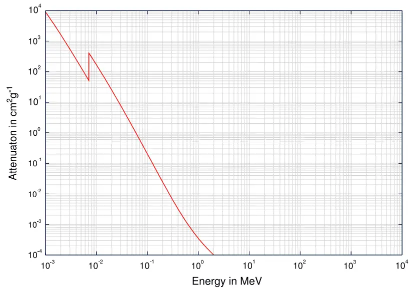
At small values of gamma ray energy the photoelectric effect dominates. The mechanism is also enhaced for materials of high atomic number Z. It is not simple to derive analytic expression for the probability of photoelectric absorption of gamma ray per atom over all ranges of gamma ray energies. The probability of photoelectric absorption per unit mass is approximately proportional to:
τ(photoelectric) = constant x ZN/E3.5
where Z is the atomic number, the exponent n varies between 4 and 5. E is the energy of the incident photon. The proportionality to higher powers of the atomic number Z is the main reason for using of high Z materials, such as lead or depleted uranium in gamma ray shields.
Although the probability of the photoelectric absorption of photon decreases, in general, with increasing photon energy, there are sharp discontinuities in the cross-section curve. These are called “absoption edges” and they correspond to the binding energies of electrons from atom’s bound shells. For photons with the energy just above the edge, the photon energy is just sufficient to undergo the photoelectric interaction with electron from bound shell, let say K-shell. The probability of such interaction is just above this edge much greater than that of photons of energy slightly below this edge. For photons below this edge the interaction with electron from K-shell in energetically impossible and therefore the probability drops abruptly. These edges occur also at binding energies of electrons from other shells (L, M, N …..).
Compton Scattering of X-rays
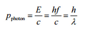
The Compton formula was published in 1923 in the Physical Review. Compton explained that the X-ray shift is caused by particle-like momentum of photons. Compton scattering formula is the mathematical relationship between the shift in wavelength and the scattering angle of the X-rays. In the case of Compton scattering the photon of frequency f collides with an electron at rest. Upon collision, the photon bounces off electron, giving up some of its initial energy (given by Planck’s formula E=hf), While the electron gains momentum (mass x velocity), the photon cannot lower its velocity. As a result of momentum conservation law, the photon must lower its momentum given by:
In Compton scattering, the incident gamma-ray photon is deflected through an angle Θ with respect to its original direction. This deflection results in a decrease in energy (decrease in photon’s frequency) of the photon and is called the Compton effect. Source: hyperphysics.phy-astr.gsu.edu
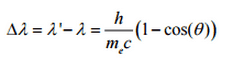
So the decrease in photon’s momentum must be translated into decrease in frequency (increase in wavelength Δλ = λ’ – λ). The shift of the wavelength increased with scattering angle according to the Compton formula:
where λ is the initial wavelength of photon λ’ is the wavelength after scattering, h is the Planck constant = 6.626 x 10⁻³⁴ J.s, me is the electron rest mass (0.511 MeV)c is the speed of light Θ is the scattering angle. The minimum change in wavelength (λ′ − λ) for the photon occurs when Θ = 0° (cos(Θ)=1) and is at least zero. The maximum change in wavelength (λ′ − λ) for the photon occurs when Θ = 180° (cos(Θ)=-1). In this case the photon transfers to the electron as much momentum as possible. The maximum change in wavelength can be derived from Compton formula:
The quantity h/mec is known as the Compton wavelength of the electron and is equal to 2.43×10⁻¹²m.
Rayleigh Scattering – Thomson Scattering
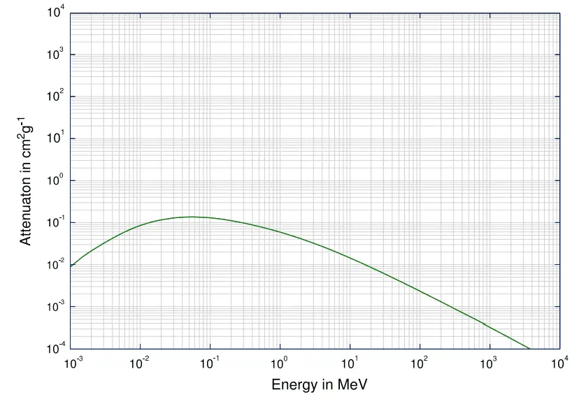
Rayleigh scattering, also known as Thomson scattering is the low-energy limit of Compton scattering. The particle kinetic energy and photon frequency do not change as a result of the scattering. Rayleigh scattering occurs as a result of an interaction between an incoming photon and an electron, the binding energy of which is significantly greater than that of the incoming photon. The incident radiation is assumed to set the electron into forced resonant oscillation such that the electron re-emits radiation of the same frequency but in all directions. In this case, the electric field of the incident wave (photon) accelerates the charged particle, causing it, in turn, to emit radiation at the same frequency as the incident wave, and thus the wave is scattered. Rayleigh scattering is significant up to ≈ 20keV and like Thomson scattering, is elastic. The total scattering cross section becomes a combination of the Rayleigh and Compton bound scattering cross sections. Thomson scattering is an important phenomenon in plasma physics and was first explained by the physicist J. J. Thomson. This interaction has great significance in the area of X-ray crystallography.
Robert Reed Burn, Introduction to Nuclear Reactor Operation, 1988.
U.S. Department of Energy, Nuclear Physics and Reactor Theory. DOE Fundamentals Handbook, Volume 1 and 2. January 1993.
Paul Reuss, Neutron Physics. EDP Sciences, 2008. ISBN: 978-2759800414.
See also:
X-rays
We hope, this article, Interaction of X-rays with Matter, helps you. If so, give us a like in the sidebar. Main purpose of this website is to help the public to learn some interesting and important information about radiation and dosimeters.
X-Ray Spectrum – Characteristic X-ray spectrum and Continuous X-ray spectrum (bremsstrahlung). For X-rays generated by X-ray tube, the part of energy that is transformed into radiation varies from zero up to the maximum energy of the electron when it hits the anode. Radiation Dosimetry
X-rays, also known as X-radiation, refers to electromagnetic radiation (no rest mass, no charge) of high energies. X-rays are high-energy photons with short wavelengths and thus very high frequency. The radiation frequency is key parameter of all photons, because it determines the energy of a photon. Photons are categorized according to the energies from low-energy radio waves and infrared radiation, through visible light, to high-energy X-rays and gamma rays.
Most X-rays have a wavelength ranging from 0.01 to 10 nanometers (3×10¹⁶ Hz to 3×10¹⁹ Hz), corresponding to energies in the range 100 eV to 100 keV. X-ray wavelengths are shorter than those of UV rays and typically longer than those of gamma rays.
X-Ray Spectrum – Characteristic and Continuous
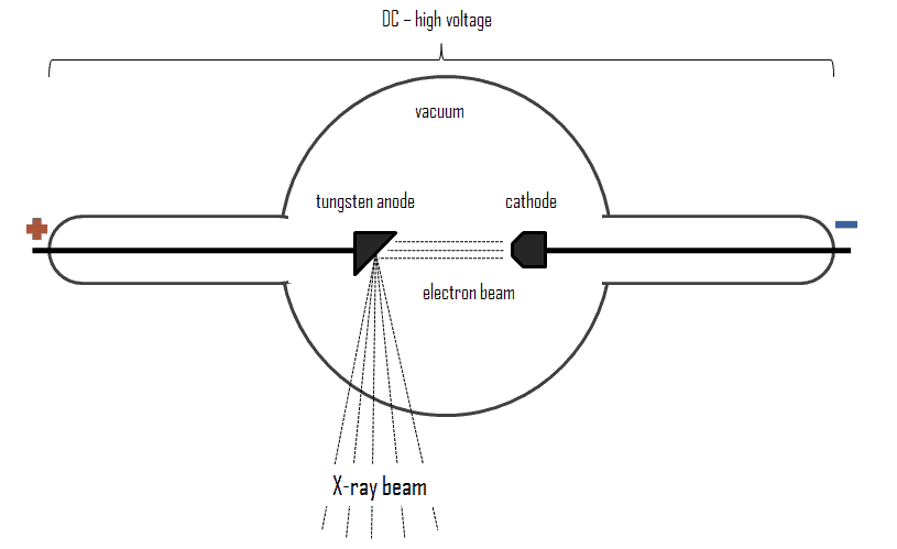
For X-rays generated by X-ray tube, the part of energy that is transformed into radiation varies from zero up to the maximum energy of the electron when it hits the anode. The maximum energy of the produced X-ray photon is limited by the energy of the incident electron, which is equal to the voltage on the tube times the electron charge, so an 100 kV tube cannot create X-rays with an energy greater than 100 keV. When the electrons hit the target, X-rays are created by two different atomic processes:
Bremsstrahlung. The bremsstrahlung is electromagnetic radiation produced by the acceleration or deceleration of an electron when deflected by strong electromagnetic fields of target high-Z (proton number) nuclei. The name bremsstrahlung comes from the German. The literal translation is ‘braking radiation’. From classical theory, when a charged particle is accelerated or decelerated, it must radiate energy. The bremsstrahlung is one of possible interactions of light charged particles with matter (especially with high atomic numbers). These X-rays have a continuous spectrum. The intensity of the X-rays increases linearly with decreasing frequency, from zero at the energy of the incident electrons, the voltage on the X-ray tube. Changing the material from which the target in the tube is made has no effect on the spectrum of this continuous radiation. If we were to switch from a molybdenum target to a copper target, for example, all features of the x-ray spectrum would change except the cutoff wavelength.
Characteristic X-ray emission. If the electron has enough energy it can knock an orbital electron out of the inner electron shell of a metal atom. Since the process leaves a vacancy in the electron energy level from which the electron came, the outer electrons of the atom cascade down to fill the lower atomic levels, and one or more characteristic X-rays are usually emitted. As a result, sharp intensity peaks appear in the spectrum at wavelengths that are a characteristic of the material from which the anode target is made. The frequencies of the characteristic X-rays can be predicted from the Bohr model.
Characteristic X-rays and Internal Conversion
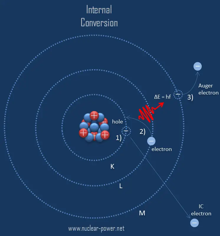
Internal conversion followed by Auger electron emission.
Internal conversion is an electromagnetic process, by which a nuclear excited state decays by the direct emission of one of its atomic electrons.
Since the process leaves a vacancy in the electron energy level from which the electron came, the outer electrons of the atom cascade down to fill the lower atomic levels, and one or more characteristic X-rays are usually emitted. Sometimes X-ray may interact with another orbital electron, which may be ejected from the atom. This second ejected electron is called an Auger electron. This is very similar to electron capture, but in case of electron capture, a nucleus changes its atomic number. As a result, the atom thus emits primary high energy electron, characteristic X-rays or secondary Auger electron, none of which originate in that nucleus.
Robert Reed Burn, Introduction to Nuclear Reactor Operation, 1988.
U.S. Department of Energy, Nuclear Physics and Reactor Theory. DOE Fundamentals Handbook, Volume 1 and 2. January 1993.
Paul Reuss, Neutron Physics. EDP Sciences, 2008. ISBN: 978-2759800414.
See also:
X-rays
We hope, this article, X-Ray Spectrum – Characteristic and Continuous, helps you. If so, give us a like in the sidebar. Main purpose of this website is to help the public to learn some interesting and important information about radiation and dosimeters.
In the photoelectric effect, X-ray undergoes an interaction with an electron which is bound in an atom.The photoelectric absorbtion dominates at low-energies of X-rays. Radiation Dosimetry
Photoelectric Absorption of X-rays
Gamma absorption by an atom. Source: laradioactivite.com/
In the photoelectric effect, a photon undergoes an interaction with an electron which is bound in an atom. In this interaction the incident photon completely disappears and an energetic photoelectron is ejected by the atom from one of its bound shells. The kinetic energy of the ejected photoelectron (Ee) is equal to the incident photon energy (hν) minus the binding energy of the photoelectron in its original shell (Eb).
Ee=hν-Eb
Therefore photoelectrons are only emitted by the photoelectric effect if photon reaches or exceeds a threshold energy – the binding energy of the electron – the work function of the material. For very high X-rays with energies of more than hundreds keV, the photoelectron carries off the majority of the incident photon energy – hν.
At small values of gamma ray energy the photoelectric effect dominates. The mechanism is also enhaced for materials of high atomic number Z. It is not simple to derive analytic expression for the probability of photoelectric absorption of gamma ray per atom over all ranges of gamma ray energies. The probability of photoelectric absorption per unit mass is approximately proportional to:
τ(photoelectric) = constant x ZN/E3.5
where Z is the atomic number, the exponent n varies between 4 and 5. E is the energy of the incident photon. The proportionality to higher powers of the atomic number Z is the main reason for using of high Z materials, such as lead or depleted uranium in gamma ray shields.
Although the probability of the photoelectric absorption of photon decreases, in general, with increasing photon energy, there are sharp discontinuities in the cross-section curve. These are called “absoption edges” and they correspond to the binding energies of electrons from atom’s bound shells. For photons with the energy just above the edge, the photon energy is just sufficient to undergo the photoelectric interaction with electron from bound shell, let say K-shell. The probability of such interaction is just above this edge much greater than that of photons of energy slightly below this edge. For photons below this edge the interaction with electron from K-shell in energetically impossible and therefore the probability drops abruptly. These edges occur also at binding energies of electrons from other shells (L, M, N …..).
Interaction of X-rays with Matter
Although a large number of possible interactions are known, there are three key interaction mechanisms with matter. The strength of these interactions depends on the energy of the X-rays and the elemental composition of the material, but not much on chemical properties, since the X-ray photon energy is much higher than chemical binding energies. The photoelectric absorbtion dominates at low-energies of X-rays, while Compton scattering dominates at higher energies.
Robert Reed Burn, Introduction to Nuclear Reactor Operation, 1988.
U.S. Department of Energy, Nuclear Physics and Reactor Theory. DOE Fundamentals Handbook, Volume 1 and 2. January 1993.
Paul Reuss, Neutron Physics. EDP Sciences, 2008. ISBN: 978-2759800414.
See also:
X-rays
We hope, this article, Photoelectric Absorption of X-rays, helps you. If so, give us a like in the sidebar. Main purpose of this website is to help the public to learn some interesting and important information about radiation and dosimeters.