What is Ionization Energy. Ionization energy, also called ionization potential, is the energy necessary to remove an electron from the neutral atom. Radiation Dosimetry
Ionization Energy
Ionization energy, also called ionization potential, is the energy necessary to remove an electron from the neutral atom.
X + energy → X+ + e−
where X is any atom or molecule capable of being ionized, X+ is that atom or molecule with an electron removed (positive ion), and e− is the removed electron.
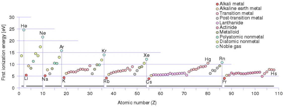
There is an ionization energy for each successive electron removed. The electrons that circle the nucleus move in fairly well-defined orbits. Some of these electrons are more tightly bound in the atom than others. For example, only 7.38 eV is required to remove the outermost electron from a lead atom, while 88,000 eV is required to remove the innermost electron.
Ionization energy is lowest for the alkali metals which have a single electron outside a closed shell.
Ionization energy increases across a row on the periodic maximum for the noble gases which have closed shells.
For example, sodium requires only 496 kJ/mol or 5.14 eV/atom to ionize it. On the other hand neon, the noble gas, immediately preceding it in the periodic table, requires 2081 kJ/mol or 21.56 eV/atom.
The ionization energy associated with removal of the first electron is most commonly used. The nth ionization energy refers to the amount of energy required to remove an electron from the species with a charge of (n-1).
1st ionization energy
X → X+ + e−
2nd ionization energy
X+ → X2+ + e−
3rd ionization energy
X2+ → X3+ + e−
For example, only 7.38 eV is required to remove the outermost electron from a lead atom, while 88,000 eV is required to remove the innermost electron.
Source: wikipedia.org License: CC BY-SA 3.0
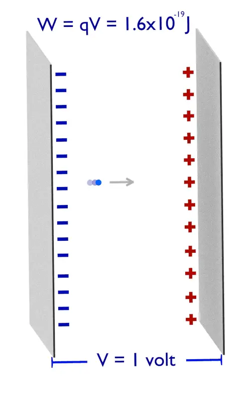
Electronvolt – Energy Unit
Electronvolt is equal to energy gained by a single electron when it is accelerated through 1 volt of electric potential difference. The work done on the charge is given by the charge times the voltage difference, therefore the work W on electron is: W = qV = (1.6 x 10⁻¹⁹ C) x (1 J/C) = 1.6 x 10⁻¹⁹ J.
Electronvolt (unit: eV). Electronvolts are a traditional unit of energy particularly in atomic and nuclear physics. Electronvolt is equal to energy gained by a single electron when it is accelerated through 1 volt of electric potential difference. The work done on the charge is given by the charge times the voltage difference, therefore the work W on electron is: W = qV = (1.6 x 10⁻¹⁹ C) x (1 J/C) = 1.6 x 10⁻¹⁹ J. Since this is very small unit, it is more convenient to use multiples of electronvolts: kilo-electronvolts (keV), mega-electronvolts (MeV), giga-electronvolts (GeV) and so on. Since Albert Einstein showed that mass and energy are equivalent and convertible one into the other, the electronvolt is also a unit of mass. It is common in particle physics, where units of mass and energy are often interchanged, to express mass in units of eV/c2, where c is the speed of light in vacuum (from E = mc2). For example, it can be said the proton has mass of 938.3 MeV, although strictly speaking it should be 938.3 MeV/c2. For another example, an electron–positron annihilation occurs when a negatively charged electron and a positively charged positron (each with a mass of 0.511 MeV/c2) collide. When an electron and a positron collide, they annihilate resulting in the complete conversion of their rest mass to pure energy (according to the E=mc2 formula) in the form of two oppositely directed 0.511 MeV gamma rays (photons).
e− + e+ → γ + γ (2x 0.511 MeV)
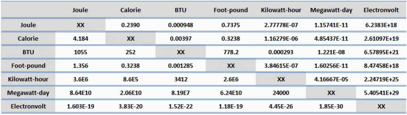
1 eV = 1.603 x 10⁻¹⁹ J
1 eV = 3.83 x 10⁻²⁰ cal
1 eV = 1.52 x 10⁻²² BTU
Example of Energies in Electronvolts
Thermal neutrons are neutrons in thermal equilibrium with a surrounding medium of temperature 290K (17 °C or 62 °F). Most probable energy at 17°C (62°F) for Maxwellian distribution is 0.025 eV (~2 km/s).
Thermal energy of a molecule is at room temperature about 0.04 eV.
Approximately 1 eV corresponds to an infrared photon of wavelength 1240 nm.
Visible light photons have energies in range 1.65 eV (red) – 3.26 eV (violet).
The first resonance in n + 238U reaction is at 6.67 eV (energy of incident neutron), which corresponds to the first virtual level in 239U, has a total width of only 0.027 eV, and the mean life of this state is 2.4×10⁻¹⁴s.
Ionization energy of atomic hydrogen is 13.6 eV.
Carbon-14 decays into nitrogen-14 through beta decay (pure beta decay). The emitted beta particles have a maximum energy of 156 keV, while their weighted mean energy is 49 keV.
High energy diagnostic medical x-ray photons have kinetic energies of about 200 keV.
Thallium 208, which is one of nuclides in the 232U decay chain, emits gamma rays of 2.6 MeV which are very energetic and highly penetrating.
Typical kinetic energy of alpha particle from radioactive decay is about 5 MeV. It is caused by the mechanism of their production.
The total energy released in a reactor is about 210 MeV per 235U fission, distributed as shown in the table. In a reactor, the average recoverable energy per fission is about 200 MeV, being the total energy minus the energy of the energy of antineutrinos that are radiated away.
Cosmic ray can have energies of 1 MeV – 1000 TeV.
References:
Reactor Physics and Thermal Hydraulics:
J. R. Lamarsh, Introduction to Nuclear Reactor Theory, 2nd ed., Addison-Wesley, Reading, MA (1983).
J. R. Lamarsh, A. J. Baratta, Introduction to Nuclear Engineering, 3d ed., Prentice-Hall, 2001, ISBN: 0-201-82498-1.
W. M. Stacey, Nuclear Reactor Physics, John Wiley & Sons, 2001, ISBN: 0- 471-39127-1.
Todreas Neil E., Kazimi Mujid S. Nuclear Systems Volume I: Thermal Hydraulic Fundamentals, Second Edition. CRC Press; 2 edition, 2012, ISBN: 978-0415802871
Zohuri B., McDaniel P. Thermodynamics in Nuclear Power Plant Systems. Springer; 2015, ISBN: 978-3-319-13419-2
Moran Michal J., Shapiro Howard N. Fundamentals of Engineering Thermodynamics, Fifth Edition, John Wiley & Sons, 2006, ISBN: 978-0-470-03037-0
Kleinstreuer C. Modern Fluid Dynamics. Springer, 2010, ISBN 978-1-4020-8670-0.
U.S. Department of Energy, THERMODYNAMICS, HEAT TRANSFER, AND FLUID FLOW. DOE Fundamentals Handbook, Volume 1, 2 and 3. June 1992.
See also:
Energy
We hope, this article, Ionization Energy, helps you. If so, give us a like in the sidebar. Main purpose of this website is to help the public to learn some interesting and important information about radiation and dosimeters.
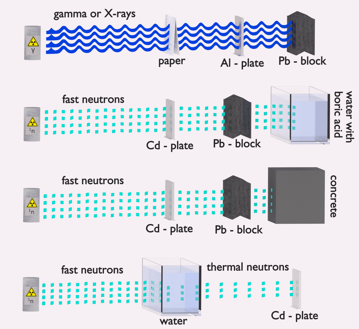
Although water is neither high density nor high Z material, it is commonly used as gamma shields. Its disadvantages are compensated with increased thickness. Radiation Dosimetry
Water as a gamma radiation shielding
In short, effective shielding of gamma radiation is in most cases based on use of materials with two following material properties:
high-density of material.
high atomic number of material (high Z materials)
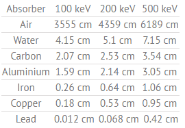
Table of Half Value Layers (in cm) for a different materials at gamma ray energies of 100, 200 and 500 keV.
Although water is neitherhigh densitynorhigh Z material, it is commonly used as gamma shields. Water provides a radiation shielding of fuel assemblies in a spent fuel pool during storage or during transports from and into the reactor core. Although water is a low-density material and low Z material, it is commonly used in nuclear power plants, because these disadvantages can be compensated with increased thickness.
Half Value Layer of Water
The half value layer expresses the thickness of absorbing material needed for reduction of the incident radiation intensity by a factor of two.
Table of Half Value Layers (in cm) for a different materials at gamma ray energies of 100, 200 and 500 keV.
References:
Reactor Physics and Thermal Hydraulics:
J. R. Lamarsh, Introduction to Nuclear Reactor Theory, 2nd ed., Addison-Wesley, Reading, MA (1983).
J. R. Lamarsh, A. J. Baratta, Introduction to Nuclear Engineering, 3d ed., Prentice-Hall, 2001, ISBN: 0-201-82498-1.
W. M. Stacey, Nuclear Reactor Physics, John Wiley & Sons, 2001, ISBN: 0- 471-39127-1.
Todreas Neil E., Kazimi Mujid S. Nuclear Systems Volume I: Thermal Hydraulic Fundamentals, Second Edition. CRC Press; 2 edition, 2012, ISBN: 978-0415802871
Zohuri B., McDaniel P. Thermodynamics in Nuclear Power Plant Systems. Springer; 2015, ISBN: 978-3-319-13419-2
Moran Michal J., Shapiro Howard N. Fundamentals of Engineering Thermodynamics, Fifth Edition, John Wiley & Sons, 2006, ISBN: 978-0-470-03037-0
Kleinstreuer C. Modern Fluid Dynamics. Springer, 2010, ISBN 978-1-4020-8670-0.
U.S. Department of Energy, THERMODYNAMICS, HEAT TRANSFER, AND FLUID FLOW. DOE Fundamentals Handbook, Volume 1, 2 and 3. June 1992.
See also:
Water
We hope, this article, Water as a gamma radiation shielding, helps you. If so, give us a like in the sidebar. Main purpose of this website is to help the public to learn some interesting and important information about radiation and dosimeters.
Water as Neutron Shielding. Water due to the high hydrogen content and the availability is efective and common neutron shielding. Radiation Dosimetry
Water as a neutron shielding
Water as a neutron shield
Water due to the high hydrogen content and the availability is efective and common neutron shielding. However, due to the low atomic number of hydrogen and oxygen, water is not acceptable shield against the gamma rays. On the other hand in some cases this disadvantage (low density) can be compensated by high thickness of the water shield. In case of neutrons, water perfectly moderates neutrons, but with absorption of neutrons by hydrogen nucleus secondary gamma rays with the high energy are produced. These gamma rays highly penetrates matter and therefore it can increase requirements on the thickness of the water shield. Adding a boric acid can help with this problem (neutron absorbtion on boron nuclei without strong gamma emission), but results in another problems with corrosion of construction materials.
Todreas Neil E., Kazimi Mujid S. Nuclear Systems Volume I: Thermal Hydraulic Fundamentals, Second Edition. CRC Press; 2 edition, 2012, ISBN: 978-0415802871
Zohuri B., McDaniel P. Thermodynamics in Nuclear Power Plant Systems. Springer; 2015, ISBN: 978-3-319-13419-2
Moran Michal J., Shapiro Howard N. Fundamentals of Engineering Thermodynamics, Fifth Edition, John Wiley & Sons, 2006, ISBN: 978-0-470-03037-0
Kleinstreuer C. Modern Fluid Dynamics. Springer, 2010, ISBN 978-1-4020-8670-0.
U.S. Department of Energy, THERMODYNAMICS, HEAT TRANSFER, AND FLUID FLOW. DOE Fundamentals Handbook, Volume 1, 2 and 3. June 1992.
See also:
Water
We hope, this article, Water as Neutron Shielding, helps you. If so, give us a like in the sidebar. Main purpose of this website is to help the public to learn some interesting and important information about radiation and dosimeters.
The personnel neutron dosimetry continues to be one of the problems in the field of radiation protection, as no single method provides the combination of energy response, sensitivity, orientation dependence characteristics
Generally every type of neutron detector must be equipped with converter and one of the conventional radiation detectors. Source: large.stanford.edu
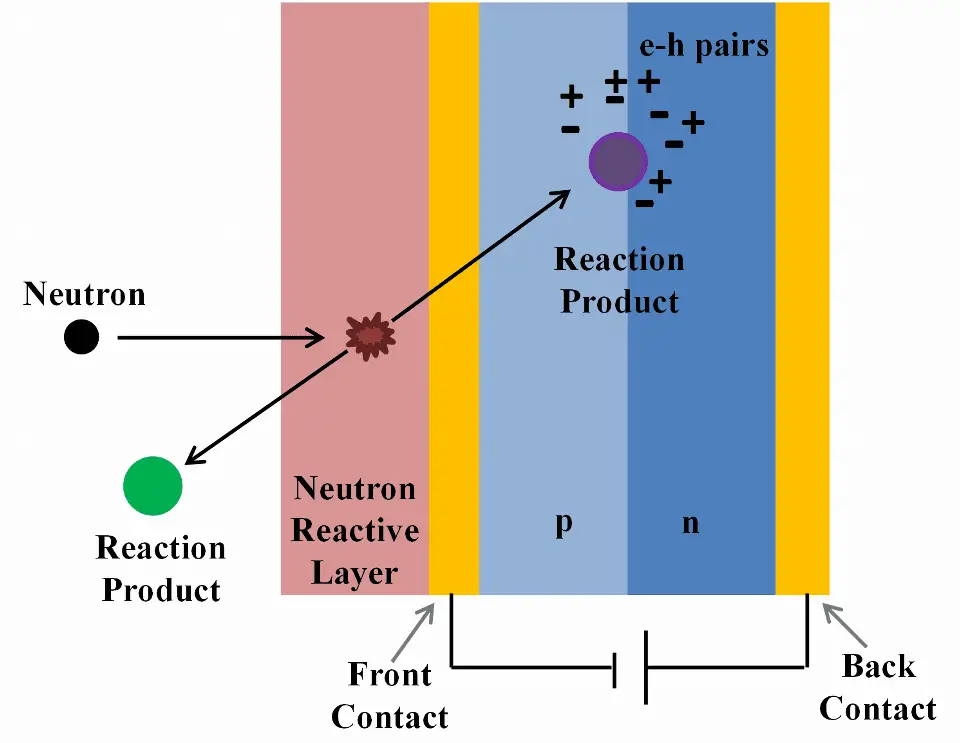
Neutron dosimetry is very specific, since the neutrons are electrically neutral particles, thus they are mainly subject to strong nuclear forces but not to electric forces. Therefore neutrons are not directly ionizing and they have usually to be converted into charged particles before they can be detected. Generally every type of neutron detector must be equipped with converter (to convert neutron radiation to common detectable radiation) and one of the conventional radiation detectors (scintillation detector, gaseous detector, semiconductor detector, etc.).
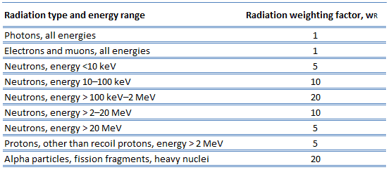
Studies have shown that alpha and neutron radiation cause greater biological damage for a given energy deposition per kg of tissue than gamma radiation does. It was discovered, biological effects of any radiation increases with the linear energy transfer (LET). In short, the biological damage from high-LET radiation (alpha particles, protons or neutrons) is much greater than that from low-LET radiation (gamma rays). This is because the living tissue can more easily repair damage from radiation that is spread over a large area than that which is concentrated in a small area. Because more biological damage is caused for the same physical dose (i.e., the same energy deposited per unit mass of tissue), one gray of alpha or neutron radiation is more harmful than one gray of gamma radiation. This fact that radiations of different types (and energies) give different biological effects for the same absorbed dose is described in terms of factors known as the relative biological effectiveness (RBE) and the radiation weighting factor (wR).
Radiation Weighting Factors – ICRP
For photon and electron radiation, the radiation weighting factorhas the value 1 independently of the energy of the radiation and for alpha radiation the value 20. For neutron radiation, the value is energy-dependent and amounts to 5 to 20.
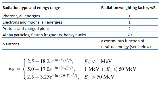
In 2007 ICRP published a new set of radiation weighting factors(ICRP Publ. 103: The 2007 Recommendations of the International Commission on Radiological Protection). These factors are given below.
Source: ICRP Publ. 103: The 2007 Recommendations of the International Commission on Radiological Protection
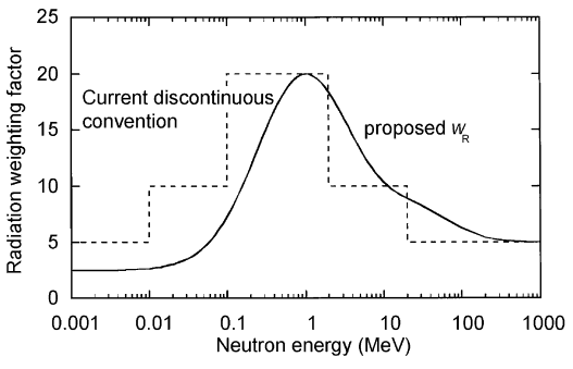
As shown in the table, a wR of 1 is for all low-LET radiations, i.e. X-rays and gamma rays of all energies as well as electrons and muons. A smooth curve, considered an approximation, was fitted to the wRvalues as a function of incident neutron energy. Note that En is the neutron energy in MeV.
The radiation weighting factor wR for neutrons introduced in Publication 60 (ICRP, 1991) as a discontinuous function of the neutron energy(- – -) and the proposed modification (—).
Thus for example, an absorbed dose of 1 Gy by alpha particles will lead to an equivalent dose of 20 Sv, and an equivalent dose of radiation is estimated to have the same biological effect as an equal amount of absorbed dose of gamma rays, which is given a weighting factor of 1.
Detection of Thermal Neutrons
Thermal neutrons are neutrons in thermal equilibrium with a surrounding medium of temperature 290K (17 °C or 62 °F). Most probable energy at 17°C (62°F) for Maxwellian distribution is 0.025 eV (~2 km/s). This part of neutron’s energy spectrum constitutes most important part of spectrum in thermal reactors.
Thermal neutrons have a different and often much larger effective neutron absorption cross-section (fission or radiative capture) for a given nuclide than fast neutrons.
In general, there are many detection principles and many types of detectors. In nuclear reactors, gaseous ionization detectors are the most common, since they are very efficient, reliable and cover a wide range of neutron flux. Various types of gaseous ionization detectors constitute so called the excore nuclear instrumentation system (NIS). The excore nuclear instrumentation system monitors the power level of the reactor by detecting neutron leakage from the reactor core.
Detection of Neutrons using Ionization Chamber
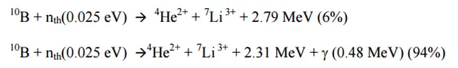
Ionization chambers are often used as the charged particle detection device. For example, if the inner surface of the ionization chamber is coated with a thin coat of boron, the (n,alpha) reaction can take place. Most of (n,alpha) reactions of thermal neutrons are 10B(n,alpha)7Li reactions accompanied by 0.48 MeV
Moreover, isotope boron-10 has high (n,alpha) reaction cross-section along the entire neutron energy spectrum. The alpha particle causes ionization within the chamber, and ejected electrons cause further secondary ionizations.
Another method for detecting neutrons using an ionization chamber is to use the gas boron trifluoride (BF3) instead of air in the chamber. The incoming neutrons produce alpha particles when they react with the boron atoms in the detector gas. Either method may be used to detect neutrons in nuclear reactor. It must be noted, BF3 counters are usually operated in the proportional region.
Detection of Fast Neutrons
Fast neutrons are neutrons of kinetic energy greater than 1 MeV (~15 000 km/s). In nuclear reactors, these neutrons are usually named fission neutrons. The fission neutrons have a Maxwell-Boltzmann distribution of energy with a mean energy (for 235U fission) 2 MeV. Inside a nuclear reactor the fast neutrons are slowed down to the thermal energies via a process called neutron moderation. These neutrons are also produced by nuclear processes such as nuclear fission or (ɑ,n) reactions.
In general, there are many detection principles and many types of detectors. Bu it must be added, detection of fast neutrons is very sophisticated discipline, since fast neutrons cross section are much smaller than in the energy range for slow neutrons. Fast neutrons are often detected by first moderating (slowing) them to thermal energies. However, during that process the information on the original energy of the neutron, its direction of travel, and the time of emission is lost.
Proton Recoil – Recoil Detectors
The most important type of detectors for fast neutrons are those which directly detect recoil particles, in particular recoil protons resulting from elastic (n, p) scattering. In fact, only hydrogen and helium nuclei are light enough for practical application. In the latter case the recoil particles are detected in a detector. Neutrons can transfer more energy to light nuclei. This method is appropriate for detecting fast neutrons allowing detection of fast neutrons without a moderator. This methods allows the energy of the neutron to be measured together with the neutron fluence, i.e. the detector can be used as a spectrometer. Typical fast neutron detectors are liquid scintillators, helium-4 based noble gas detectors and plastic detectors (scintillators). For example, the plastic has a high hydrogen content, therefore, it is useful for fast neutron detectors, when used as a scintillator.
Bonner Spheres Spectrometer
There are several methods for detecting slow neutrons, and few methods for detecting fast neutrons. Therefore, one technique for measuring fast neutrons is to convert them to slow
neutrons, and then measure the slow neutrons. One of possible methods is based on Bonner spheres. The method was first described in 1960 by Ewing and Tom W. Bonner and employs thermal neutron detectors (usually inorganic scintillators such as 6LiI) embedded in moderating spheres of different sizes. Bonner spheres have been used widely for the measurement of neutron spectra with neutron energies ranged from thermal up to at least 20 MeV. A Bonner sphere neutron spectrometer (BSS) consists of a thermal-neutron detector, a set polyethylene spherical shells and two optional lead shells of various sizes. In order to detect thermal neutrons a 3He detector or inorganic scintillators such as 6LiI can be used. LiGlass scintillators are very popular for detection of thermal neutrons. The advantage of LiGlass scintillators is their stability and their large range of sizes.
Detection of Neutrons using Scintillation Counter
Scintillation counters are used to measure radiation in a variety of applications including hand held radiation survey meters, personnel and environmental monitoring for radioactive contamination, medical imaging, radiometric assay, nuclear security and nuclear plant safety. They are widely used because they can be made inexpensively yet with good efficiency, and can measure both the intensity and the energy of incident radiation.
Neutrons. Since the neutrons are electrically neutral particles, they are mainly subject to strong nuclear forces but not to electric forces. Therefore neutrons are not directly ionizing and they have usually to be converted into charged particles before they can be detected. Generally every type of neutron detector must be equipped with converter (to convert neutron radiation to common detectable radiation) and one of the conventional radiation detectors (scintillation detector, gaseous detector, semiconductor detector, etc.). Fast neutrons (>0.5 MeV) primarily rely on the recoil proton in (n,p) reactions. Materials rich in hydrogen, for example plastic scintillators, are therefore best suited for their detection. Thermal neutrons rely on nuclear reactions such as the (n,γ) or (n,α) reactions, to produce ionization. Materials such as LiI(Eu) or glass silicates are therefore particularly well-suited for the detection of thermal neutrons. The advantage of 6LiGlass scintillators is their stability and their large range of sizes.
Neutron Thermoluminescent Dosimeter – Neutron TLD
The personnel neutron dosimetry continues to be one of the problems in the field of radiation protection, as no single method provides the combination of energy response, sensitivity, orientation dependence characteristics and accuracy necessary to meet the needs of a personnel dosimeter.
The most commonly used personnel neutron dosimeters for radiation protection purposes are thermoluminescent dosimeters and albedo dosimeters. Both are based on this phenomenon – thermoluminescence. For this purpose, lithium fluoride (LiF) as sensitive material (chip) is widely used. Lithium fluoride TLD is used for gamma and neutron exposure (indirectly, using the Li-6 (n,alpha)) nuclear reaction. Small crystals of LiF (lithium fluoride) are the most common TLD dosimeters since they have the same absorption properties as soft tissue. Lithium has two stable isotopes, lithium-6 (7.4 %) and lithium-7 (92.6 %). Li-6 is the isotope sensitive to neutrons. In order to record neutrons, LiF crystal dosimeters may be enriched in lithium-6 to enhance the lithium-6 (n,alpha) nuclear reaction. The efficiency of the detector depends on the energy of the neutrons. Because the interaction of neutrons with any element is highly dependent on energy, making a dosimeter independent of the energy of neutrons is very difficult. In order to separate thermal neutrons and photons, LiF dosimeters are mostly utilized, containing different percentage of lithium-6. LiF chip enriched in lithium-6, which is very sensitive to thermal neutrons and LiF chip containing very little of lithium-6, which has a negligible neutron response.
The principle of neutron TLDs is then similar as for gamma radiation TLDs. In the LiF chip, there are impurities (e.g. manganese or magnesium), which produce trap states for energetic electrons. The impurity causes traps in the crystalline lattice where, following irradiation (to alpha radiation), electrons are held. When the crystal is warmed, the trapped electrons are released and light is emitted. The amount of light is related to the dose of radiation received by the crystal.
Thermoluminescent Albedo Neutron Dosimeter
Albedo neutron dosimetry is based on the effect of moderation and backscattering of neutrons by the human body. Albedo, the latin word for “whiteness”, was defined by Lambert as the fraction of the incident light reflected diffusely by a surface. Moderation and backscattering of neutrons by the human body creates a neutron flux at the body surface in the thermal and intermediate energy range. These backscattered neutrons called albedo neutrons, can be detected by a dosimeter (usually a LiF TLD chip), placed on the body which is designed to detect thermal neutrons. Albedo dosimeters have been found to be the only dosimeters which can measure doses due to neutrons over the whole range of energies. Usually, two types of lithium fluoride are used to separate doses contributed by gamma-rays and neutrons. LiF chip enriched in lithium-6, which is very sensitive to thermal neutrons and LiF chip containing very little of lithium-6, which has a negligible neutron response.
See also:
Personal Dosimetry
We hope, this article, Neutron Dosimetry – Neutron Dosimeter, helps you. If so, give us a like in the sidebar. Main purpose of this website is to help the public to learn some interesting and important information about radiation and dosimeters.
X-ray dosimetry is very specific, because high-energy photons interact differently with matter. Geiger counters may be used to detect gamma radiation and X-rays (thin-walled tubes) collectively known as photons. Radiation Dosimetry
X-ray dosimetry is very specific, because high-energy photons interact differently with matter. High-energy photons can travel thousands of feet in air and can easily pass through various materials. Moreover, high-energy photons can ionize atoms indirectly and directly (despite they are electrically neutral) through the photoelectric effect and the Compton effect. But secondary (indirect) ionization is much more significant.
Detectors of X-Rays
Detectors may be also categorized according to sensitive materials and methods that can be utilized to make a measurement:
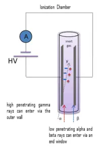
Gamma rays have very little trouble in penetrating the metal walls of the chamber. Therefore, ionization chambers may be used to detect gamma radiation and X-rays collectively known as photons, and for this the windowless tube is used. Ionization chambers have a good uniform response to radiation over a wide range of energies and are the preferred means of measuring high levels of gamma radiation. Some problems are caused by the fact, that alpha particles are more ionising than beta particles and than gamma rays, so more current is produced in the ionization chamber region by alpha than beta and gamma. Gamma rays deposit significantly lower amount of energy to the detector than other particles.
Detection of X-Rays using Geiger Counter
Detector of Ionizing Radiation – Geiger Tube
Geiger counter can detect ionizing radiation such as alpha and beta particles, neutrons, X-rays and gamma rays using the ionization effect produced in a Geiger–Müller tube, which gives its name to the instrument. The voltage of detector is adjusted so that the conditions correspond to the Geiger-Mueller region.
The high amplification factor of the Geiger counter is the major advantage over the ionization chamber. Geiger counter is therefore a much more sensitive device than other chambers. It is often used in the detection of low-level gamma rays and beta particles for this reason.
Windowless type
Gamma rays have very little trouble in penetrating the metal walls of the chamber. Therefore, Geiger counters may be used to detect gamma radiation and X-rays (thin-walled tubes) collectively known as photons, and for this the windowless tube is used.
A thick walled tube is used for gamma radiation detection above energies of about 25 KeV, this type generally has an overall wall thickness of about 1-2 mm of chrome steel.
A thin walled tube is used for low energy photons (X-rays or gamma rays) and high energy beta particles. The transition from thin walled to thick walled design takes place at the 300–400 keV energy levels. Above these levels thick walled designs are used, and beneath these levels the direct gas ionisation effect is predominant.
Detection of X-rays using Scintillation Counter
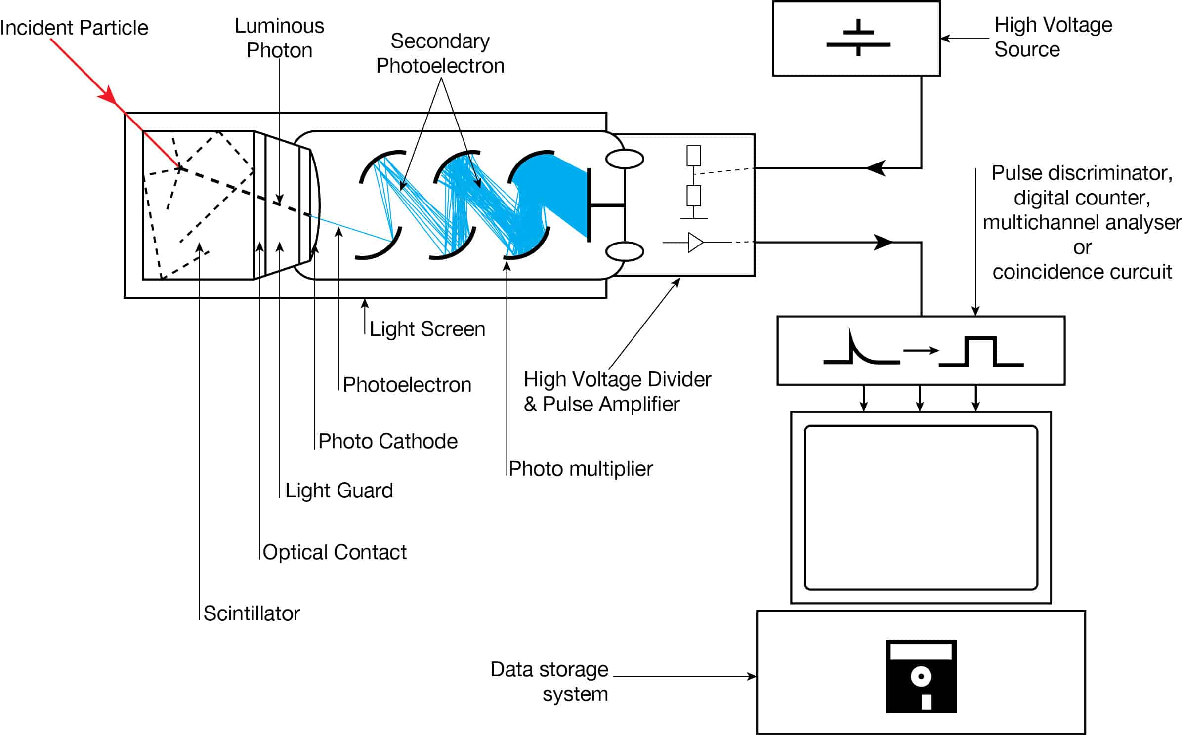
Apparatus with a scintillating crystal, photomultiplier, and data acquisition components. Source: wikipedia.org License CC BY-SA 3.0
Scintillation counters are used to measure radiation in a variety of applications including hand held radiation survey meters, personnel and environmental monitoring for radioactive contamination, medical imaging, radiometric assay, nuclear security and nuclear plant safety. They are widely used because they can be made inexpensively yet with good efficiency, and can measure both the intensity and the energy of incident radiation.
X-Rays.High-Z materials are best suited as scintillators for the detection of gamma rays. The most widely used scintillation material is NaI(Tl) (thallium-doped sodium iodide). The iodine provides most of the stopping power in sodium iodide (since it has a high Z = 53). These crystalline scintillators are characterized by high density, high atomic number, and pulse decay times of approximately 1 microsecond (~ 10-6 sec). Scintillation in inorganic crystals is typically slower than in organic ones. They exhibit high efficiency for detection of gamma rays and are capable of handling high count rates. Inorganic crystals can be cut to small sizes and arranged in an array configuration so as to provide position sensitivity. This feature is widely used in medical imaging to detect X-rays or gamma rays. Inorganic scintillators are better at detecting gamma rays and X-rays. This is due to their high density and atomic number which gives a high electron density.
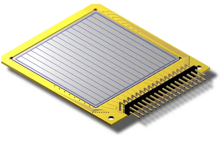
Detection of X-Rays using Semiconductors – HPGe Detectors
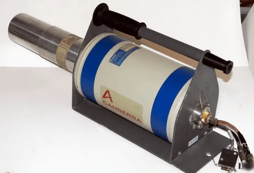
HPGe detector with LN2 cryostat Source: canberra.com
As was written, the study and analysis of gamma ray spectra for scientific and technical use is called gamma spectroscopy, and gamma ray spectrometers are the instruments which observe and collect such data. A gamma ray spectrometer (GRS) is a sophisticated device for measuring the energy distribution of gamma radiation. For the measurement of gamma rays above several hundred keV, there are two detector categories of major importance, inorganic scintillators as NaI(Tl) and semiconductor detectors. If a perfect energy resolution is required, we have to use germanium-based detector, such as the HPGe detector. Germanium-based semiconductor detectors are most commonly used where a very good energy resolution is required, especially for gamma spectroscopy, as well as x-ray spectroscopy. In gamma spectroscopy, germanium is preferred due to its atomic number being much higher than silicon and which increases the probability of gamma ray interaction. Moreover, germanium has lower average energy necessary to create an electron-hole pair, which is 3.6 eV for silicon and 2.9 eV for germanium. This also provides the latter a better resolution in energy. The FWHM (full width at half maximum) for germanium detectors is a function of energy. For a 1.3 MeV photon, the FWHM is 2.1 keV, which is very low.
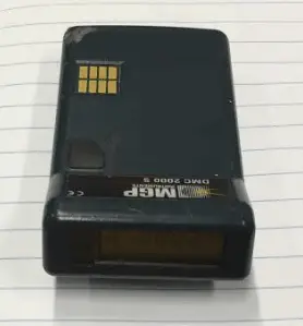
EPD – Electronic Personal Dosimeter
EPD – Electronic Personal Dosimeters with Si chip
An electronic personal dosimeter is modern dosimeter, which can give a continuous readout of cumulative dose and current dose rate, and can warn the person wearing it when a specified dose rate or a cumulative dose is exceeded. EPDs are especially useful in high dose areas where residence time of the wearer is limited due to dose constraints.
Characteristics of EPDs
The electronic personal dosimeter, EPD, is able to display a direct reading of the detected dose or dose rate in real time. Electronic dosimeters may be used as a supplemental dosimeter as well a primary dosimeter. The passive dosimeters and the electronic personal dosimeters are often used together to complement each other. To estimate effective doses, dosimeters must be worn on a position of the body representative of its exposure, typically between the waist and the neck, on the front of the torso, facing the radioactive source. Dosimeters are usually worn on the outside of clothing, around the chest or torso to represent dose to the “whole body”. Dosimeters may also be worn on the extremities or near the eye to measure equivalent dose to these tissues.
The dosimeter can be reset, usually after taking a reading for record purposes, and thereby re-used multiple times. The EPDs have a top mounted display to make them easy to read when they are clipped to your breast pocket. The digital display gives both dose and dose rateinformation usually in mSv and mSv/h. The EPD has a dose rate alarm, and a dose alarm. These alarms are programmable. Different alarms can be set for different activities.
For example:
dose rate alarm at 100 μSv/h,
dose alarm: 100 μSv.
If an alarm set point is reached, the relevant display flashes along with a red light, and quite a piercing noise is generated. You can clear the dose rate alarm by retreating to a lower radiation field, but you cannot clear the dose alarm until you get to a EPD reader. EPDs can also give a bleep for every 1 or 10 μSv they register. This gives you an audible indication of the radiation fields. Some EPDs have wireless communication capabilities. EPDs are capable of measuring a wide radiation dose range from routine (μSv) levels to emergency levels (hundreds mSv or units of Sieverts) with high precision, and may display the exposure rate as well as accumulated exposure values. Of the dosimeter technologies, electronic personal dosimeters are generally the most expensive, largest in size, and the most versatile.
Robert Reed Burn, Introduction to Nuclear Reactor Operation, 1988.
U.S. Department of Energy, Nuclear Physics and Reactor Theory. DOE Fundamentals Handbook, Volume 1 and 2. January 1993.
Paul Reuss, Neutron Physics. EDP Sciences, 2008. ISBN: 978-2759800414.
See also:
Personal Dosimetry
We hope, this article, X-Ray Dosimetry – X-Ray Dosimeter, helps you. If so, give us a like in the sidebar. Main purpose of this website is to help the public to learn some interesting and important information about radiation and dosimeters.
Gamma dosimetry is the measurement, calculation and assessment of the absorbed doses and assigning those doses to individuals. Radiation Dosimetry
Gamma dosimetry is the measurement, calculation and assessment of the absorbed doses and assigning those doses to individuals. It is the science and practice that attempts to quantitatively relate specific measures made in a radiation field to chemical and/or biological changes that the radiation would produce in a target.
Since there are two types of radiation exposure, external and internal exposure, dosimetry may be also categorized as:
External Dosimetry. External exposure is radiation that comes from outside our body and interacts with us. In this case, we analyze predominantly exposure from gamma rays and beta particles, since alpha particles, in general, constitute no external exposure hazard because the particles generally do not pass through skin. Since photons and beta interact through electromagnetic forces and neutrons interact through nuclear forces, their detection methods and dosimetry are substantially different. The source of radiation can be, for example, a piece of equipment that produces the radiation like a container with a radioactive materials, or like an x-ray machine. External dosimetry is based on measurements with a dosimeter, or inferred from measurements made by other radiological protection instruments.
Internal Dosimetry. If the source of radiation is inside our body, we say, it is internal exposure. The intake of radioactive material can occur through various pathways such as ingestion of radioactive contamination in food or liquids. Protection from internal exposure is more complicated. Most radionuclides will give you much more radiation dose if they can somehow enter your body, than they would if they remained outside. Internal dosimetry assessment relies on a variety of monitoring, bio-assay or radiation imaging techniques.
Studies have shown that alpha and neutron radiation cause greater biological damage for a given energy deposition per kg of tissue than gamma radiation does. It was discovered, biological effects of any radiation increases with the linear energy transfer (LET). In short, the biological damage from high-LET radiation (alpha particles, protons or neutrons) is much greater than that from low-LET radiation (gamma rays). This is because the living tissue can more easily repair damage from radiation that is spread over a large area than that which is concentrated in a small area. Because more biological damage is caused for the same physical dose (i.e., the same energy deposited per unit mass of tissue), one gray of alpha or neutron radiation is more harmful than one gray of gamma radiation. This fact that radiations of different types (and energies) give different biological effects for the same absorbed dose is described in terms of factors known as the relative biological effectiveness (RBE) and the radiation weighting factor (wR).
Radiation Weighting Factors – ICRP
For photon and electron radiation, the radiation weighting factorhas the value 1 independently of the energy of the radiation and for alpha radiation the value 20. For neutron radiation, the value is energy-dependent and amounts to 5 to 20.
In 2007 ICRP published a new set of radiation weighting factors(ICRP Publ. 103: The 2007 Recommendations of the International Commission on Radiological Protection). These factors are given below.
Source: ICRP Publ. 103: The 2007 Recommendations of the International Commission on Radiological Protection
As shown in the table, a wR of 1 is for all low-LET radiations, i.e. X-rays and gamma rays of all energies as well as electrons and muons. A smooth curve, considered an approximation, was fitted to the wRvalues as a function of incident neutron energy. Note that En is the neutron energy in MeV.
The radiation weighting factor wR for neutrons introduced in Publication 60 (ICRP, 1991) as a discontinuous function of the neutron energy(- – -) and the proposed modification (—).
Thus for example, an absorbed dose of 1 Gy by alpha particles will lead to an equivalent dose of 20 Sv, and an equivalent dose of radiation is estimated to have the same biological effect as an equal amount of absorbed dose of gamma rays, which is given a weighting factor of 1.
Detectors of Gamma Radiation
Detectors may be also categorized according to sensitive materials and methods that can be utilized to make a measurement:
Detection of Gamma Radiation using Ionization Chamber
Gamma rays have very little trouble in penetrating the metal walls of the chamber. Therefore, ionization chambers may be used to detect gamma radiation and X-rays collectively known as photons, and for this the windowless tube is used. Ionization chambers have a good uniform response to radiation over a wide range of energies and are the preferred means of measuring high levels of gamma radiation. Some problems are caused by the fact, that alpha particles are more ionising than beta particles and than gamma rays, so more current is produced in the ionization chamber region by alpha than beta and gamma. Gamma rays deposit significantly lower amount of energy to the detector than other particles.
The efficiency of the chamber can be further increased by the use of a high pressure gas. Typically a pressure of 8-10 atmospheres can be used, and various noble gases are employed. For example, high-pressure xenon (HPXe) ionization chambers are ideal for use in uncontrolled environments, as a detector’s response has been shown to be uniform over large temperature ranges (20–170°C). The higher pressure results in a greater gas density and thereby a greater chance of collision with the fill gas and ion-pair creation by incident gamma radiation. Because of the increased wall thickness required to withstand this high pressure, only gamma radiation can be detected. These detectors are used in survey meters and for environmental monitoring.
Detection of Gamma Radiation using Geiger Counter
Detector of Ionizing Radiation – Geiger Tube
Geiger counter can detect ionizing radiation such as alpha and beta particles, neutrons, and gamma rays using the ionization effect produced in a Geiger–Müller tube, which gives its name to the instrument. The voltage of detector is adjusted so that the conditions correspond to the Geiger-Mueller region.
The high amplification factor of the Geiger counter is the major advantage over the ionization chamber. Geiger counter is therefore a much more sensitive device than other chambers. It is often used in the detection of low-level gamma rays and beta particles for this reason.
Detection of Gamma Radiation using Scintillation Counter
Apparatus with a scintillating crystal, photomultiplier, and data acquisition components. Source: wikipedia.org License CC BY-SA 3.0
Scintillation counters are used to measure radiation in a variety of applications including hand held radiation survey meters, personnel and environmental monitoring for radioactive contamination, medical imaging, radiometric assay, nuclear security and nuclear plant safety. They are widely used because they can be made inexpensively yet with good efficiency, and can measure both the intensity and the energy of incident radiation.
Gamma Rays. High-Z materials are best suited as scintillators for the detection of gamma rays. The most widely used scintillation material is NaI(Tl) (thallium-doped sodium iodide). The iodine provides most of the stopping power in sodium iodide (since it has a high Z = 53). These crystalline scintillators are characterized by high density, high atomic number, and pulse decay times of approximately 1 microsecond (~ 10-6 sec). Scintillation in inorganic crystals is typically slower than in organic ones. They exhibit high efficiency for detection of gamma rays and are capable of handling high count rates. Inorganic crystals can be cut to small sizes and arranged in an array configuration so as to provide position sensitivity. This feature is widely used in medical imaging to detect X-rays or gamma rays. Inorganic scintillators are better at detecting gamma rays and X-rays. This is due to their high density and atomic number which gives a high electron density.
Detection of Gamma Radiation using Semiconductors – HPGe Detectors
HPGe detector with LN2 cryostat Source: canberra.com
As was written, the study and analysis of gamma ray spectra for scientific and technical use is called gamma spectroscopy, and gamma ray spectrometers are the instruments which observe and collect such data. A gamma ray spectrometer (GRS) is a sophisticated device for measuring the energy distribution of gamma radiation. For the measurement of gamma rays above several hundred keV, there are two detector categories of major importance, inorganic scintillators as NaI(Tl) and semiconductor detectors. If a perfect energy resolution is required, we have to use germanium-based detector, such as the HPGe detector. Germanium-based semiconductor detectors are most commonly used where a very good energy resolution is required, especially for gamma spectroscopy, as well as x-ray spectroscopy. In gamma spectroscopy, germanium is preferred due to its atomic number being much higher than silicon and which increases the probability of gamma ray interaction. Moreover, germanium has lower average energy necessary to create an electron-hole pair, which is 3.6 eV for silicon and 2.9 eV for germanium. This also provides the latter a better resolution in energy. The FWHM (full width at half maximum) for germanium detectors is a function of energy. For a 1.3 MeV photon, the FWHM is 2.1 keV, which is very low.
EPD – Electronic Personal Dosimeter
EPD – Electronic Personal Dosimeters with Si chip
An electronic personal dosimeter is modern dosimeter, which can give a continuous readout of cumulative dose and current dose rate, and can warn the person wearing it when a specified dose rate or a cumulative dose is exceeded. EPDs are especially useful in high dose areas where residence time of the wearer is limited due to dose constraints.
Characteristics of EPDs
The electronic personal dosimeter, EPD, is able to display a direct reading of the detected dose or dose rate in real time. Electronic dosimeters may be used as a supplemental dosimeter as well a primary dosimeter. The passive dosimeters and the electronic personal dosimeters are often used together to complement each other. To estimate effective doses, dosimeters must be worn on a position of the body representative of its exposure, typically between the waist and the neck, on the front of the torso, facing the radioactive source. Dosimeters are usually worn on the outside of clothing, around the chest or torso to represent dose to the “whole body”. Dosimeters may also be worn on the extremities or near the eye to measure equivalent dose to these tissues.
The dosimeter can be reset, usually after taking a reading for record purposes, and thereby re-used multiple times. The EPDs have a top mounted display to make them easy to read when they are clipped to your breast pocket. The digital display gives both dose and dose rateinformation usually in mSv and mSv/h. The EPD has a dose rate alarm, and a dose alarm. These alarms are programmable. Different alarms can be set for different activities.
For example:
dose rate alarm at 100 μSv/h,
dose alarm: 100 μSv.
If an alarm set point is reached, the relevant display flashes along with a red light, and quite a piercing noise is generated. You can clear the dose rate alarm by retreating to a lower radiation field, but you cannot clear the dose alarm until you get to a EPD reader. EPDs can also give a bleep for every 1 or 10 μSv they register. This gives you an audible indication of the radiation fields. Some EPDs have wireless communication capabilities. EPDs are capable of measuring a wide radiation dose range from routine (μSv) levels to emergency levels (hundreds mSv or units of Sieverts) with high precision, and may display the exposure rate as well as accumulated exposure values. Of the dosimeter technologies, electronic personal dosimeters are generally the most expensive, largest in size, and the most versatile.
Robert Reed Burn, Introduction to Nuclear Reactor Operation, 1988.
U.S. Department of Energy, Nuclear Physics and Reactor Theory. DOE Fundamentals Handbook, Volume 1 and 2. January 1993.
Paul Reuss, Neutron Physics. EDP Sciences, 2008. ISBN: 978-2759800414.
See also:
Personal Dosimetry
We hope, this article, Gamma Dosimetry – Gamma Dosimeter, helps you. If so, give us a like in the sidebar. Main purpose of this website is to help the public to learn some interesting and important information about radiation and dosimeters.
Beta dosimetry is very specific, because beta particles are more penetrating than alpha particles. The film badge can be used to measure and record radiation exposure due to gamma rays, X-rays and beta particles. Radiation Dosimetry
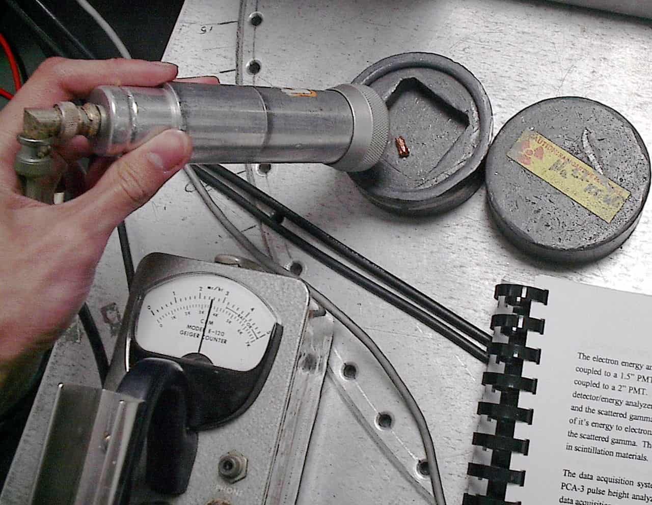
Laboratory use of a Geiger counter with end-window probe to measure beta radiation Source: wikipedia.org License: Public Domain
Beta dosimetry is very specific, because beta particles are more penetrating than alpha particles. On the other hand a thin aluminum plate can stop them.
Alpha and beta particles, in general, constitute no external exposure hazard because the particles generally do not pass through skin. On the other hand, alpha and beta radiation is very harmful, when their radionuclides are ingested or inhaled. Internal exposure is more dangerous than external exposure, since we are carrying the source of radiation inside our bodies and we cannot use any of radiation protection principles (time, distance, shielding).
Studies have shown that alpha and neutron radiation cause greater biological damage for a given energy deposition per kg of tissue than gamma radiation does. It was discovered, biological effects of any radiation increases with the linear energy transfer (LET). In short, the biological damage from high-LET radiation (alpha particles, protons or neutrons) is much greater than that from low-LET radiation (gamma rays). This is because the living tissue can more easily repair damage from radiation that is spread over a large area than that which is concentrated in a small area. Because more biological damage is caused for the same physical dose (i.e., the same energy deposited per unit mass of tissue), one gray of alpha or neutron radiation is more harmful than one gray of gamma radiation. This fact that radiations of different types (and energies) give different biological effects for the same absorbed dose is described in terms of factors known as the relative biological effectiveness (RBE) and the radiation weighting factor (wR).
Radiation Weighting Factors – ICRP
For photon and electron radiation, the radiation weighting factorhas the value 1 independently of the energy of the radiation and for alpha radiation the value 20. For neutron radiation, the value is energy-dependent and amounts to 5 to 20.
In 2007 ICRP published a new set of radiation weighting factors(ICRP Publ. 103: The 2007 Recommendations of the International Commission on Radiological Protection). These factors are given below.
Source: ICRP Publ. 103: The 2007 Recommendations of the International Commission on Radiological Protection
As shown in the table, a wR of 1 is for all low-LET radiations, i.e. X-rays and gamma rays of all energies as well as electrons and muons. A smooth curve, considered an approximation, was fitted to the wRvalues as a function of incident neutron energy. Note that En is the neutron energy in MeV.
The radiation weighting factor wR for neutrons introduced in Publication 60 (ICRP, 1991) as a discontinuous function of the neutron energy(- – -) and the proposed modification (—).
Thus for example, an absorbed dose of 1 Gy by alpha particles will lead to an equivalent dose of 20 Sv, and an equivalent dose of radiation is estimated to have the same biological effect as an equal amount of absorbed dose of gamma rays, which is given a weighting factor of 1.
Detectors of Beta Radiation
Detectors may be also categorized according to sensitive materials and methods that can be utilized to make a measurement:
Detection of Beta Radiation using Ionization Chamber
For alpha and beta particles to be detected by ionization chambers, they must be provided with a thin window. This “end-window” must be thin enough for the alpha and beta particles to penetrate. However, a window of almost any thickness will prevent an alpha particle from entering the chamber. The window is usually made of mica with a density of about 1.5 – 2.0 mg/cm2.
Ionization chamber may be, for example, used for the measurement of tritium in the air. These devices are known as tritium-in-air monitors. Tritium is a radioactive isotope, but it emits a very weak form of radiation, a low-energy beta particle that is similar to an electron. It is a pure beta emitter (i.e. beta emitter without an accompanying gamma radiation). The electron’s kinetic energy varies, with an average of 5.7 keV, while the remaining energy is carried off by the nearly undetectable electron antineutrino. Such a very low energy of electron causes, that the electron cannot penetrate the skin or even does not travel very far in air. Beta particles from tritium can penetrate only about 6.0 mm of air. It is practically impossible to design a detector whose walls these beta particles can penetrate. Instead, tritium-in-air monitor pumps the tritium-contaminated air right through an ionization chamber, so that all of the energy of the beta particles can be usefully converted to producing ion pairs inside the chamber.
Detection of Beta Radiation using Scintillation Counter
Scintillation counters are used to measure radiation in a variety of applications including hand held radiation survey meters, personnel and environmental monitoring for radioactive contamination, medical imaging, radiometric assay, nuclear security and nuclear plant safety. They are widely used because they can be made inexpensively yet with good efficiency, and can measure both the intensity and the energy of incident radiation.
Beta Particles. For detection of beta particles, organic scintillators can be used. Pure organic crystals include crystals of anthracene, stilbene and naphthalene. The decay time of this type of phosphor is approximately 10 nanoseconds. This type of crystal is frequently used in the detection of beta particles. Organic scintillators, having a lower Z than inorganic crystals, are best suited for the detection of low-energy (< 10 MeV) beta particles.
Detection of Beta Radiation using Semiconductors – Silicon Strip Detectors
Silicon-based detectors are very good for tracking charged particles. A silicon strip detector is an arrangement of strip like shaped implants acting as charge collecting electrodes.
Silicon strip detectors 5 x 5 cm2 in area are quite common and are used in series (just like planes of MWPCs) to determine charged-particle trajectories to position-accuracies of the order of several μm in the transverse direction. Placed on a low doped fully depleted silicon wafer these implants form a one-dimensional array of diodes. By connecting each of the metalized strips to a charge sensitive amplifier a position sensitive detector is built. Two dimensional position measurements can be achieved by applying an additional strip like doping on the wafer backside by use of a double sided technology. Such devices can be used to measure small impact parameters and thereby determine whether some charged particle originated from a primary collision or was the decay product of a primary particle that traveled a small distance from the original interaction, and then decayed.
Portable Survey Meters
Portable survey meters are radiation detectors used by radiological technicians to measure ambient dose rate. These portable instruments usually have rate meters. In nuclear facilities, these portable survey meters are typically used by radiation protection technicians, which are responsible for following operations in the field to help assure that radiation protection policies are carried out and that jobs are implemented in accordance with the ALARA principle. Their responsibilities include:
Providing assistance and advice to workers to motivate them to adopt an ALARA behaviour.
Following jobs to ensure the respect of safety and radiation protection procedures.
In some plants, stopping work in case of serious deviation from dosimetric objectives, or when there is a significantly increasing radiological risk for workers.
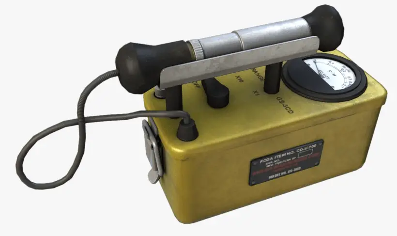
The typical radiation survey meter is, for example, the RDS-31, which is a multi-purpose radiation survey meter that uses a G-M detector. It has optional alpha, beta, and gamma external probes. It measures 3.9 x 2.6 x 1.3 inches and can be handheld, or worn by pocket, belt clip, or pouch. It has a five-digit, backlit, LCD display. Geiger counters operate at such a high voltage that the size of the output pulse is always the same, regardless of how many ion pairs were created in the detector. Geiger counters are mainly used for portable instrumentation due to its sensitivity, simple counting circuit, and ability to detect low-level radiation.
Film Badge Dosimeter
Film badge dosimeters are for one-time use only, they cannot be reused. A film badge dosimeter is dosimeter, that is worn at the surface of the body by the person being monitored, and it records of the radiation dose received. The film badge is used to measure and record radiation exposure due to gamma rays, X-rays and beta particles. The badge incorporates a series of filters (lead, tin, cadmium and plastic) to determine the quality of the radiation. To monitor beta particle emission, the filters use various densities of plastic or even label material. It is typical for a single badge to contain a series of filters of different thicknesses and of different materials; the precise choice may be determined by the environment to be monitored.
Examples of filters:
There is an open window that makes it possible for weaker radiations to reach the film.
A thin plastic filter which attenuates beta radiation but passes all other radiations
A thick plastic filter which passes all but the lowest energy photon radiation and absorbs all but the highest beta radiation.
A dural filter which progressively absorbs photon radiation at energies below 65 keV as well as beta radiation.
A tin/lead filter of a thickness which allows an energy independent dose response of the film over the photon energy range 75 keV to 2 MeV.
A cadmium lead filter can be used for thermal neutrons detection. The capture of neutrons ((n,gamma) reactions) by cadmiumproduces gamma rays which blacken the film thus enabling assessment of exposure to neutrons.
Robert Reed Burn, Introduction to Nuclear Reactor Operation, 1988.
U.S. Department of Energy, Nuclear Physics and Reactor Theory. DOE Fundamentals Handbook, Volume 1 and 2. January 1993.
Paul Reuss, Neutron Physics. EDP Sciences, 2008. ISBN: 978-2759800414.
See also:
Personal Dosimetry
We hope, this article, Beta Dosimetry – Beta Dosimeter, helps you. If so, give us a like in the sidebar. Main purpose of this website is to help the public to learn some interesting and important information about radiation and dosimeters.
Alpha dosimetry is very specific, because alpha particles travel only a few centimeters in air but deposit all their energies along their short paths. Radiation Dosimetry
Alpha dosimetry is very specific, because alpha particles travel only a few centimeters in air but deposit all their energies along their short paths, thus the amount of energy transferred is very high. Alpha and beta particles, in general, constitute no external exposure hazard because the particles generally do not pass through skin. On the other hand, alpha radiation is very harmful, when alpha radionuclides are ingested or inhaled. Internal exposure is more dangerous than external exposure, since we are carrying the source of radiation inside our bodies and we cannot use any of radiation protection principles (time, distance, shielding).
Studies have shown that alpha and neutron radiation cause greater biological damage for a given energy deposition per kg of tissue than gamma radiation does. It was discovered, biological effects of any radiation increases with the linear energy transfer (LET). In short, the biological damage from high-LET radiation (alpha particles, protons or neutrons) is much greater than that from low-LET radiation (gamma rays). This is because the living tissue can more easily repair damage from radiation that is spread over a large area than that which is concentrated in a small area. Because more biological damage is caused for the same physical dose (i.e., the same energy deposited per unit mass of tissue), one gray of alpha or neutron radiation is more harmful than one gray of gamma radiation. This fact that radiations of different types (and energies) give different biological effects for the same absorbed dose is described in terms of factors known as the relative biological effectiveness (RBE) and the radiation weighting factor (wR).
Radiation Weighting Factors – ICRP
For photon and electron radiation, the radiation weighting factorhas the value 1 independently of the energy of the radiation and for alpha radiation the value 20. For neutron radiation, the value is energy-dependent and amounts to 5 to 20.
In 2007 ICRP published a new set of radiation weighting factors(ICRP Publ. 103: The 2007 Recommendations of the International Commission on Radiological Protection). These factors are given below.
Source: ICRP Publ. 103: The 2007 Recommendations of the International Commission on Radiological Protection
As shown in the table, a wR of 1 is for all low-LET radiations, i.e. X-rays and gamma rays of all energies as well as electrons and muons. A smooth curve, considered an approximation, was fitted to the wRvalues as a function of incident neutron energy. Note that En is the neutron energy in MeV.
The radiation weighting factor wR for neutrons introduced in Publication 60 (ICRP, 1991) as a discontinuous function of the neutron energy(- – -) and the proposed modification (—).
Thus for example, an absorbed dose of 1 Gy by alpha particles will lead to an equivalent dose of 20 Sv, and an equivalent dose of radiation is estimated to have the same biological effect as an equal amount of absorbed dose of gamma rays, which is given a weighting factor of 1.
Detectors of Alpha Radiation
Detectors may be also categorized according to sensitive materials and methods that can be utilized to make a measurement:
Detection of Alpha Radiation using Ionization Chamber
For alpha and beta particles to be detected by ionization chambers, they must be provided with a thin window. This “end-window” must be thin enough for the alpha and beta particles to penetrate. However, a window of almost any thickness will prevent an alpha particle from entering the chamber. The window is usually made of mica with a density of about 1.5 – 2.0 mg/cm2. But it does not mean, alpha radiation cannot be detected by an ionization chamber.
For example, in some kind of smoke detectors, you can meet man-made radionuclides such as americium-241, which is a source of alpha particles. The smoke detector has two ionization chambers, one open to the air, and a reference chamber which does not allow the entry of particles. The radioactive source emits alpha particles into both chambers, which ionizes some air molecules. The free-air chamber allows the entry of smoke particles to the sensitive volume and to change attenuation of alpha particles. If any smoke particles enter the free-air chamber, some of the ions will attach to the particles and not be available to carry the current in that chamber. An electronic circuit detects that a current difference has developed between the open and sealed chambers, and sounds the alarm.
Detection of Alpha Radiation using Geiger-Mueller Counter
Geiger counters are mainly used for portable instrumentation due to its sensitivity, simple counting circuit, and ability to detect low-level radiation. Although the major use of Geiger counters is probably in individual particle detection, they are also found in gamma survey meters. They are able to detect almost all types of radiation, but there are slight differences in the Geiger-Mueller tube. However, the Geiger-Müller tube produces a pulse output which is the same magnitude for all detected radiation, so a Geiger counter with an end window tube cannot distinguish between alpha and beta particles.
End-Window type
For alpha and beta particles to be detected by Geiger counters, they must be provided with a thin window. This “end-window” must be thin enough for the alpha and beta particles to penetrate. However, a window of almost any thickness will prevent an alpha particle from entering the chamber. The window is usually made of mica with a density of about 1.5 – 2.0 mg/cm2 to allow low-energy beta particles (e.g. from carbon-14) to enter the detector. The efficiency reduction for alpha is due to the attenuation effect of the end window, though distance from the surface being checked also has a significant effect, and ideally a source of alpha radiation should be less than 10mm from the detector due to attenuation in air.
Detection of Alpha using Scintillation Counter
Scintillation counters are used to measure radiation in a variety of applications including hand held radiation survey meters, personnel and environmental monitoring for radioactive contamination, medical imaging, radiometric assay, nuclear security and nuclear plant safety. They are widely used because they can be made inexpensively yet with good efficiency, and can measure both the intensity and the energy of incident radiation.
Scintillation counters can be used to detect alpha, beta, gamma radiation. They can be used also for detection of neutrons. For these purposes, different scintillators are used:
Alpha Particles and Heavy Ions. Due to the very high ionizing power of heavy ions, scintillation counters are usually not ideal for the detection of heavy ions. For equal energies, a proton will produce 1/4 to 1/2 the light of an electron, while alpha particles will produce only about 1/10 the light. Where needed, inorganic crystals, e.g. CsI(Tl), ZnS(Ag) (typically used in thin sheets as α-particle monitors), should be preferred to organic materials. Pure CsI is a fast and dense scintillating material with relatively low light yield that increases significantly with cooling. The drawbacks of CsI are a high temperature gradient and a slight hygroscopicity.
Detection of Alpha using Semiconductors – Silicon Strip Detectors
Silicon-based detectors are very good for tracking charged particles. A silicon strip detector is an arrangement of strip like shaped implants acting as charge collecting electrodes.
Silicon strip detectors 5 x 5 cm2 in area are quite common and are used in series (just like planes of MWPCs) to determine charged-particle trajectories to position-accuracies of the order of several μm in the transverse direction. Placed on a low doped fully depleted silicon wafer these implants form a one-dimensional array of diodes. By connecting each of the metalized strips to a charge sensitive amplifier a position sensitive detector is built. Two dimensional position measurements can be achieved by applying an additional strip like doping on the wafer backside by use of a double sided technology. Such devices can be used to measure small impact parameters and thereby determine whether some charged particle originated from a primary collision or was the decay product of a primary particle that traveled a small distance from the original interaction, and then decayed.
Portable Survey Meter
Portable survey meters are radiation detectors used by radiological technicians to measure ambient dose rate. These portable instruments usually have rate meters. In nuclear facilities, these portable survey meters are typically used by radiation protection technicians, which are responsible for following operations in the field to help assure that radiation protection policies are carried out and that jobs are implemented in accordance with the ALARA principle. Their responsibilities include:
Providing assistance and advice to workers to motivate them to adopt an ALARA behaviour.
Following jobs to ensure the respect of safety and radiation protection procedures.
In some plants, stopping work in case of serious deviation from dosimetric objectives, or when there is a significantly increasing radiological risk for workers.
The typical radiation survey meter is, for example, the RDS-31, which is a multi-purpose radiation survey meter that uses a G-M detector. It has optional alpha, beta, and gamma external probes. It measures 3.9 x 2.6 x 1.3 inches and can be handheld, or worn by pocket, belt clip, or pouch. It has a five-digit, backlit, LCD display. Geiger counters operate at such a high voltage that the size of the output pulse is always the same, regardless of how many ion pairs were created in the detector. Geiger counters are mainly used for portable instrumentation due to its sensitivity, simple counting circuit, and ability to detect low-level radiation.
Robert Reed Burn, Introduction to Nuclear Reactor Operation, 1988.
U.S. Department of Energy, Nuclear Physics and Reactor Theory. DOE Fundamentals Handbook, Volume 1 and 2. January 1993.
Paul Reuss, Neutron Physics. EDP Sciences, 2008. ISBN: 978-2759800414.
See also:
Personal Dosimetry
We hope, this article, Alpha Dosimetry, helps you. If so, give us a like in the sidebar. Main purpose of this website is to help the public to learn some interesting and important information about radiation and dosimeters.
A whole-body counter is an instrument that measures the amounts of gamma-emitting radionuclides in the body. In nuclear facilities, whole-body counters are used for measurement of radioactivity within the human body. Radiation Dosimetry
A whole-body counter is an instrument that measures the amounts of gamma-emitting radionuclides in the body (i.e. it is a gamma spectrometer). In nuclear facilities, these counters are used for measurement of radioactivity within the human body, that means, for internal contamination measurements. This must not be confused with a “whole body monitor” which used for personnel exit monitoring, which is the term used in radiation protection for checking for external contamination of a whole body of a person leaving a radioactive contamination controlled area. Whole-body counters are very sensitive devices and therefore they are often surrounded by large quantities of lead shielding to reduce the background radiation. A whole-body counter consists, for example, of a stand-up booth with two large-area NaI scintillation detectors. The upper detector monitors lungs, the lower detector monitors the gastrointestinal tract.
It must be noted, all people also have some radioactive isotopes inside their bodies from birth. These isotopes are especially potassium-40, carbon-14 and also the isotopes of uranium and thorium. The average annual radiation dose to a person from internal radioactive materials other than radon is about 0.3 mSv/year of which:
2 mSv/year comes from potassium-40,
12 mSv/year comes from the uranium and thorium series,
12 μSv/year comes from carbon-40.
The variation in radiation dose from one person to another is not as great, but it is detected also by a whole-body counter.
Gamma Spectroscopy
HPGe detector with LN2 cryostat Source: canberra.com
If a gamma ray is emitted from a radioactive element within the human body due to radioactive decay, and its energy is sufficient to escape then it can be detected. This would be by means of gamma spectrometer. Spectroscopes, or spectrometers, are sophisticated devices designed to measure the spectral power distribution of a source. The incident radiation generates a signal that allows to determine the energy of the incident particle. Most radioactive sources produce gamma rays, which are of various energies and intensities. Gamma rays frequently accompany the emission of alpha and beta radiation. When these emissions are detected and analyzed with a spectroscopy system, a gamma-ray energy spectrum can be produced. Gamma rays from radioactive decay are in the energy range from a few keV to ~8 MeV, corresponding to the typical energy levels in nuclei with reasonably long lifetimes. As was written, they are produced by the decay of nuclei as they transition from a high energy state to a lower state. A detailed analysis of this spectrum is typically used to determine the identity and quantity of gamma emitters present in a sample, and is a vital tool in radiometric assay. The gamma spectrum is characteristic of the gamma-emitting nuclides contained in the source.
For the measurement of gamma rays above several hundred keV, there are two detector categories of major importance, inorganic scintillators as NaI(Tl) and semiconductor detectors. In the previous articles, we described the gamma spectroscopy using scintillation detector, which consists of a suitable scintillator crystal, a photomultiplier tube, and a circuit for measuring the height of the pulses produced by the photomultiplier. The advantages of a scintillation counter are its efficiency (large size and high density) and the high precision and counting rates that are possible. Due to the high atomic number of iodine, a large number of all interactions will result in complete absorption of gamma-ray energy, so the photo fraction will be high.
But if a perfect energy resolution is required, we have to use germanium-based detector, such as the HPGe detector. Germanium-based semiconductor detectors are most commonly used where a very good energy resolution is required, especially for gamma spectroscopy, as well as x-ray spectroscopy. In gamma spectroscopy, germanium is preferred due to its atomic number being much higher than silicon and which increases the probability of gamma ray interaction. Moreover, germanium has lower average energy necessary to create an electron-hole pair, which is 3.6 eV for silicon and 2.9 eV for germanium. This also provides the latter a better resolution in energy. The FWHM (full width at half maximum) for germanium detectors is a function of energy. For a 1.3 MeV photon, the FWHM is 2.1 keV, which is very low.
Internal Dose Uptake
If the source of radiation is inside our body, we say, it is internal exposure. The intake of radioactive material can occur through various pathways such as ingestion of radioactive contamination in food or liquids, inhalation of radioactive gases, or through intact or wounded skin. Most radionuclides will give you much more radiation dose if they can somehow enter your body, than they would if they remained outside. For internal doses, we first should distinguish between intake and uptake. Intake means what a person takes in. Uptake means what a person keeps.
When a radioactive compound enters the body, the activity will decrease with time, due both to radioactive decay and to biological clearance. The decrease varies from one radioactive compound to another. For this purpose, the biological half-life is defined in radiation protection.
The biological half-life is the time taken for the amount of a particular element in the body to decrease to half of its initial value due to elimination by biological processes alone, when the rate of removal is roughly exponential. The biological half-life depends on the rate at which the body normally uses a particular compound of an element. Radioactive isotopes that were ingested or taken in through other pathways will gradually be removed from the body via bowels, kidneys, respiration and perspiration. This means that a radioactive substance can be expelled before it has had the chance to decay.
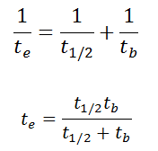
As a result, the biological half-life significantly influences the effective half-life and the overall dose from internal contamination. If a radioactive compound with radioactive half-life (t1/2) is cleared from the body with a biological half-life tb, the effective half-life (te) is given by the expression:
As can be seen, the biological mechanisms always decreases the overall dose from internal contamination. Moreover, if t1/2 is large in comparison to tb, the effective half-life is approximately the same as tb.
For example, tritium has the biological half-life about 10 days, while the radioactive half-life is about 12 years. On the other hand, radionuclides with very short radioactive half-lives have also very short effective half-lives. These radionuclides will deliver, for all practical purposes, the total radiation dose within the first few days or weeks after intake.
For tritium, the annual limit intake (ALI) is 1 x 10⁹ Bq. If you take in 1 x 10⁹ Bq of tritium, you will receive a whole-body dose of 20 mSv. The committed effective dose, E(t), is therefore 20 mSv. It does not depend whether a person intakes this amount of activity in a short time or in a long time. In every case, this person gets the same whole-body dose of 20 mSv.
Robert Reed Burn, Introduction to Nuclear Reactor Operation, 1988.
U.S. Department of Energy, Nuclear Physics and Reactor Theory. DOE Fundamentals Handbook, Volume 1 and 2. January 1993.
Paul Reuss, Neutron Physics. EDP Sciences, 2008. ISBN: 978-2759800414.
See also:
Dosimetry in NPPs
We hope, this article, Whole-Body Counter – Whole-Body Counting, helps you. If so, give us a like in the sidebar. Main purpose of this website is to help the public to learn some interesting and important information about radiation and dosimeters.
Contamination meters are instruments for surface contamination measurement. These monitors may utilize proportional counters with a large area, thin window detector similar to Hand & Shoe Monitors.
Contamination meters are instruments for surface contamination measurement. Generally, surface contamination means that radioactive material has been deposited on surfaces (such as walls, floors). It may be loosely deposited, much like ordinary dust, or it may be quite firmly fixed by chemical reaction. This distinction is important, and we classify surface contamination on the basis of how easily it can be removed. In nuclear facilities, contamination monitors are installed usually at the exit from the controlled areas. These monitors may utilize proportional counters with a large area, thin window detector similar to Hand & Shoe Monitors. When instruments are operated in the proportional region, the voltage must be kept constant. If a voltage remains constant the gas amplification factor also does not change. Proportional counter detection instruments are very sensitive to low levels of radiation. By proper functional arrangements, modifications, and biasing, the proportional counter can be used to detect alpha, beta, gamma, or neutron radiation in mixed radiation fields. The electronics sorts the alpha, the beta-gamma pulses and displays both in a bar-type display.
Robert Reed Burn, Introduction to Nuclear Reactor Operation, 1988.
U.S. Department of Energy, Nuclear Physics and Reactor Theory. DOE Fundamentals Handbook, Volume 1 and 2. January 1993.
Paul Reuss, Neutron Physics. EDP Sciences, 2008. ISBN: 978-2759800414.
See also:
Dosimetry in NPPs
We hope, this article, Alpha Beta Gamma Contamination Meter, helps you. If so, give us a like in the sidebar. Main purpose of this website is to help the public to learn some interesting and important information about radiation and dosimeters.