Facebook Instagram Youtube Twitter

What is Sievert – Gray – Becquerel – Conversion – Calculation – Definition

But what is the relationship between becquerels (radioactivity), grays (absorbed dose) and sieverts (equivalent dose)? This article shows, how to convert and calculate these quantities. Radiation Dosimetry

sievert - radiationIn radiation protection, the sievert is a derived unit of equivalent dose and effective dose. The sievert represents the equivalent biological effect of the deposit of a joule of gamma rays energy in a kilogram of human tissue. But what is the relationship between becquerels (radioactivity) and sieverts (equivalent dose)?

In previous chapters, we have discussed radioactivity and the intensity of a radioactive source, measured usually in becquerels. But any radioactive source represents no biological risk as long as it is isolated from the environments. However, when people or another system (also non-biological) are exposed to radiation, energy is deposited in the material and radiation dose is delivered.

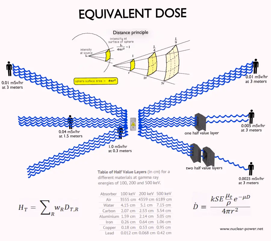
It is therefore very important to distinguish between radioactivity of a radioactive source and the radiation dose which may result from the source. Generally, the radiation dose depends on the following factors regarding to the radioactive source:

  • Activity. Activity of the source directly influences the radiation dose deposited in the material.
  • Type of radiation. Each type of radiation interacts with matter in a different way. For example charged particles with high energies can directly ionize atoms. On the other hand electrically neutral particles interacts only indirectly, but can also transfer some or all of their energies to the matter.
  • Distance. The amount of radiation exposure depends on the distance from the source of radiation. Similarly to a heat from a fire, if you are too close, the intensity of heat radiation is high and you can get burned. If you are at the right distance, you can withstand there without any problems and moreover it is comfortable. If you are too far from heat source, the insufficiency of heat can also hurt you. This analogy, in a certain sense, can be applied to radiation also from radiation sources.
  • Time. The amount of radiation exposure depends directly (linearly) on the time people spend near the source of radiation.
  • Shielding. Finally, the radiation dose also depends on the material between the source and the object. If the source is too intensive and time or distance do not provide sufficient radiation protection, the shielding can be used.

The danger of ionizing radiation lies in the fact that the radiation is invisible and not directly detectable by human senses. People can neither see nor feel radiation, yet it deposits energy to the molecules of the body. The energy is transferred in small quantities for each interaction between the radiation and a molecule and there are usually many such interactions.

Sievert and Gray

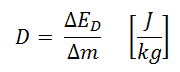
Absorbed dose is defined as the amount of energy deposited by ionizing radiation in a substance. Absorbed dose is given the symbol D.  The absorbed dose is usually measured in a unit called the gray (Gy), which is derived from the SI system. The non-SI unit rad is sometimes also used, predominantly in the USA.
absorbed dose - definition

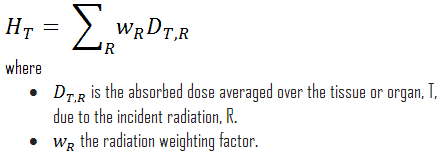
For radiation protection purposes, the absorbed dose is averaged over an organ or tissue, T, and this absorbed dose average is weighted for the radiation quality in terms of the radiation weighting factor, wR, for the type and energy of radiation incident on the body. The radiation weighting factor is a dimensionless factor used to determine the equivalent dose from the absorbed dose averaged over a tissue or organ and is based on the type of radiation absorbed. The resulting weighted dose was designated as the organ- or tissue equivalent dose:
equivalent dose - equation - definition

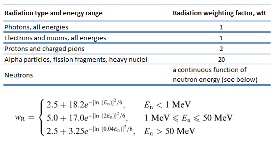
Radiation weighting factors - current - ICRP
Table of radiation weighting factors. Source: ICRP Publ. 103: The 2007 Recommendations of the International Commission on Radiological Protection

An equivalent dose of one Sievert represents that quantity of radiation dose that is equivalent, in terms of specified biological damage, to one gray of X-rays or gamma rays. A dose of one Sv caused by gamma radiation is equivalent to an energy deposition of one joule in a kilogram of a tissue. That means one sievert is equivalent to one gray of gamma rays deposited in certain tissue. On the other hand, similar biological damage (one sievert) can be caused only by 1/20 gray of alpha radiation (due to high wR of alpha radiation). Therefore, the sievert is not a physical dose unit. For example, an absorbed dose of 1 Gy by alpha particles will lead to an equivalent dose of 20 Sv. This may seem to be a paradox. It implies that the energy of the incident radiation field in joules has increased by a factor of 20, thereby violating the laws of Conservation of energy. However, this is not the case. Sievert is derived from the physical quantity absorbed dose, but also takes into account the biological effectiveness of the radiation, which is dependent on the radiation type and energy. The radiation weighting factor causes that the sievert cannot be a physical unit.

One sievert is a large amount of equivalent dose. A person who has absorbed a whole body dose of 1 Sv has absorbed one joule of energy in each kg of body tissue (in case of gamma rays).

Equivalent doses measured in industry and medicine often have usually lower doses than one sievert, and the following multiples are often used:

1 mSv (millisievert) = 1E-3 Sv

1 µSv (microsievert) = 1E-6 Sv

Conversions from the SI units to other units are as follows:

  • 1 Sv = 100 rem
  • 1 mSv = 100 mrem

Radiation Weighting Factors – ICRP

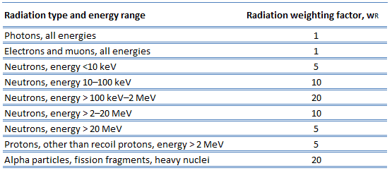
For photon and electron radiation, the radiation weighting factor has the value 1 independently of the energy of the radiation and for alpha radiation the value 20. For neutron radiation, the value is energy-dependent and amounts to 5 to 20.
Radiation weighting factors
Source: ICRP, 2003. Relative Biological Effectiveness (RBE), Quality Factor (Q), and Radiation Weighting Factor (wR). ICRP Publication 92. Ann. ICRP 33 (4).

In 2007 ICRP published a new set of radiation weighting factors (ICRP Publ. 103: The 2007 Recommendations of the International Commission on Radiological Protection). These factors are given below.

Radiation weighting factors - current - ICRP
Source: ICRP, 2007. Publ. 103: The 2007 Recommendations of the International Commission on Radiological Protection.

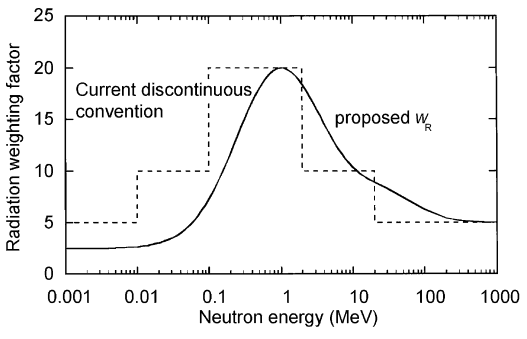
As shown in the table, a wR of 1 is for all low-LET radiations, i.e. X-rays and gamma rays of all energies as well as electrons and muons. A smooth curve, considered an approximation, was fitted to the wR values as a function of incident neutron energy. Note that En is the neutron energy in MeV.
radiation weighting factor - neutrons - ICRP
The radiation weighting factor wR for neutrons introduced in Publication 60 (ICRP, 1991) as a discontinuous function of the neutron energy(- – -) and the proposed modification (—).

Thus for example, an absorbed dose of 1 Gy by alpha particles will lead to an equivalent dose of 20 Sv, and an equivalent dose of radiation is estimated to have the same biological effect as an equal amount of absorbed dose of gamma rays, which is given a weighting factor of 1.

See also: Quality Factor

Effective Dose – Sieverts
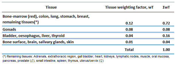
tissue weighting factor - ICRPThe effective dose is a dose quantity defined as the sum of the tissue-equivalent doses weighted by the ICRP organ (tissue) weighting factors, wT, which takes into account the varying sensitivity of different organs and tissues to radiation.

Effective dose allows to determine the biological consequences of partial irradiation (non-uniform) to consequences of complete irradiation. Various body tissues react to ionising radiation in different ways, so the ICRP has assigned sensitivity factors to specified tissues and organs so that the effect of partial irradiation can be calculated if the irradiated regions are known.

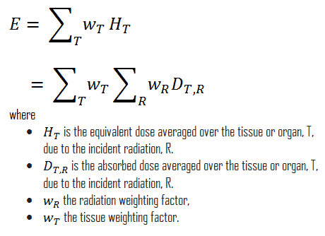
In Publication 60, the ICRP defined effective dose as the doubly weighted sum of absorbed dose in all the organs and tissues of the body. Dose limits are set in terms of effective dose and apply to the individual for radiological protection purposes, including the assessment of risk in general terms. Mathematically, the effective dose can be expressed as:
effective dose - definition


Examples of Doses in Sieverts

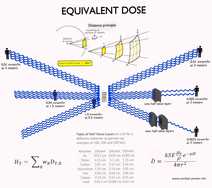
We must note that radiation is all around us. In, around, and above the world we live in. It is a natural energy force that surrounds us. It is a part of our natural world that has been here since the birth of our planet. In the following points we try to express enormous ranges of radiation exposure, which can be obtained from various sources.

  • 0.05 µSv – Sleeping next to someone
  • 0.09 µSv – Living within 30 miles of a nuclear power plant for a year
  • 0.1 µSv – Eating one banana
  • 0.3 µSv – Living within 50 miles of a coal power plant for a year
  • 10 µSv – Average daily dose received from natural background
  • 20 µSv – Chest X-ray
  • 40 µSv – A 5-hour airplane flight
  • 600 µSv – mammogram
  • 1 000 µSv – Dose limit for individual members of the public, total effective dose per annum
  • 3 650 µSv – Average yearly dose received from natural background
  • 5 800 µSv – Chest CT scan
  • 10 000 µSv – Average yearly dose received from natural background in Ramsar, Iran
  • 20 000 µSv – single full-body CT scan
  • 175 000 µSv – Annual dose from natural radiation on a monazite beach near Guarapari, Brazil.
  • 5 000 000 µSv – Dose that kills a human with a 50% risk within 30 days (LD50/30), if the dose is received over a very short duration.

As can be seen, low-level doses are common for everyday life. The previous examples can help illustrate relative magnitudes. From biological consequences point of view, it is very important to distinguish between doses received over short and extended periods.  An “acute dose” is one that occurs over a short and finite period of time, while a “chronic dose” is a dose that continues for an extended period of time so that it is better described by a dose rate. High doses tend to kill cells, while low doses tend to damage or change them. Low doses spread out over long periods of time don’t cause an immediate problem to any body organ. The effects of low doses of radiation occur at the level of the cell, and the results may not be observed for many years.

Calculation of Shielded Dose Rate in Sieverts

Assume the point isotropic source which contains 1.0 Ci of 137Cs, which has a half-life of 30.2 years. Note that the relationship between half-life and the amount of a radionuclide required to give an activity of one curie is shown below. This amount of material can be calculated using λ, which is the decay constant of certain nuclide:

Curie - Unit of Activity

About 94.6 percent decays by beta emission to a metastable nuclear isomer of barium: barium-137m. The main photon peak of Ba-137m is 662 keV. For this calculation, assume that all decays go through this channel.

Calculate the primary photon dose rate, in gray per hour (Gy.h-1), at the outer surface of a 5 cm thick lead shield. Then calculate the equivalent dose rate. Assume that this external radiation field penetrate uniformly through the whole body. Primary photon dose rate neglects all secondary particles. Assume that the effective distance of the source from the dose point is 10 cm. We shall also assume that the dose point is soft tissue and it can reasonably be simulated by water and we use the mass energy absorption coefficient for water.

See also: Gamma Ray Attenuation

See also: Shielding of Gamma Rays

Solution:

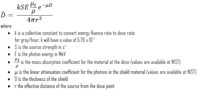
The primary photon dose rate is attenuated exponentially, and the dose rate from primary photons, taking account of the shield, is given by:
dose rate calculation

As can be seen, we do not account for the buildup of secondary radiation. If secondary particles are produced or if the primary radiation changes its energy or direction, then the effective attenuation will be much less.  This assumption generally underestimates the true dose rate, especially for thick shields and when the dose point is close to the shield surface, but this assumption simplifies all calculations. For this case the true dose rate (with the buildup of secondary radiation) will be more than two times higher.

To calculate the absorbed dose rate, we have to use in the formula:

  • k = 5.76 x 10⁻⁷
  • S = 3.7 x 10¹⁰ s-1
  • E = 0.662 MeV
  • μt/ρ =  0.0326 cm2/g (values are available at NIST)
  • μ =  1.289 cm-1 (values are available at NIST)
  • D = 5 cm
  • r = 10 cm

Result:

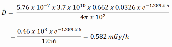
The resulting absorbed dose rate in grays per hour is then:
absorbed dose rate - gray - calculation

Since the radiation weighting factor for gamma rays is equal to one and we have assumed the uniform radiation field, we can directly calculate the equivalent dose rate from the absorbed dose rate as:

equivalent dose - sievert - calculation

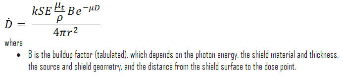
If we want to account for the buildup of secondary radiation, then we have to include the buildup factor. The extended formula for the dose rate is then:
absorbed dose rate - gray


——–

References:

Radiation Protection:

  1. Knoll, Glenn F., Radiation Detection and Measurement 4th Edition, Wiley, 8/2010. ISBN-13: 978-0470131480.
  2. Stabin, Michael G., Radiation Protection and Dosimetry: An Introduction to Health Physics, Springer, 10/2010. ISBN-13: 978-1441923912.
  3. Martin, James E., Physics for Radiation Protection 3rd Edition, Wiley-VCH, 4/2013. ISBN-13: 978-3527411764.
  4. U.S.NRC, NUCLEAR REACTOR CONCEPTS
  5. U.S. Department of Energy, Nuclear Physics and Reactor Theory. DOE Fundamentals Handbook, Volume 1 and 2. January 1993.

Nuclear and Reactor Physics:

  1. J. R. Lamarsh, Introduction to Nuclear Reactor Theory, 2nd ed., Addison-Wesley, Reading, MA (1983).
  2. J. R. Lamarsh, A. J. Baratta, Introduction to Nuclear Engineering, 3d ed., Prentice-Hall, 2001, ISBN: 0-201-82498-1.
  3. W. M. Stacey, Nuclear Reactor Physics, John Wiley & Sons, 2001, ISBN: 0- 471-39127-1.
  4. Glasstone, Sesonske. Nuclear Reactor Engineering: Reactor Systems Engineering, Springer; 4th edition, 1994, ISBN: 978-0412985317
  5. W.S.C. Williams. Nuclear and Particle Physics. Clarendon Press; 1 edition, 1991, ISBN: 978-0198520467
  6. G.R.Keepin. Physics of Nuclear Kinetics. Addison-Wesley Pub. Co; 1st edition, 1965
  7. Robert Reed Burn, Introduction to Nuclear Reactor Operation, 1988.
  8. U.S. Department of Energy, Nuclear Physics and Reactor Theory. DOE Fundamentals Handbook, Volume 1 and 2. January 1993.
  9. Paul Reuss, Neutron Physics. EDP Sciences, 2008. ISBN: 978-2759800414.

See also:

Sievert

We hope, this article, Sievert – Gray – Becquerel – Conversion – Calculation, helps you. If so, give us a like in the sidebar. Main purpose of this website is to help the public to learn some interesting and important information about radiation and dosimeters.
What is Example of Doses in Sieverts – Definition

Examples of Doses in Sieverts. In the following points we try to express enormous ranges of radiation exposure in sieverts, which can be obtained from various sources. Radiation Dosimetry

sievert - radiationIn radiation protection, the sievert is a derived unit of equivalent dose and effective dose. The sievert represents the equivalent biological effect of the deposit of a joule of gamma rays energy in a kilogram of human tissue. Unit of sievert is of importance in radiation protection and was named after the Swedish scientist Rolf Sievert, who did a lot of the early work on dosimetry in radiation therapy.

One sievert is a large amount of equivalent dose. A person who has absorbed a whole body dose of 1 Sv has absorbed one joule of energy in each kg of body tissue (in case of gamma rays).

Equivalent doses measured in industry and medicine often have usually lower doses than one sievert, and the following multiples are often used:

1 mSv (millisievert) = 1E-3 Sv

1 µSv (microsievert) = 1E-6 Sv

Conversions from the SI units to other units are as follows:

  • 1 Sv = 100 rem
  • 1 mSv = 100 mrem

Examples of Doses in Sieverts

We must note that radiation is all around us. In, around, and above the world we live in. It is a natural energy force that surrounds us. It is a part of our natural world that has been here since the birth of our planet. In the following points we try to express enormous ranges of radiation exposure, which can be obtained from various sources.

  • 0.05 µSv – Sleeping next to someone
  • 0.09 µSv – Living within 30 miles of a nuclear power plant for a year
  • 0.1 µSv – Eating one banana
  • 0.3 µSv – Living within 50 miles of a coal power plant for a year
  • 10 µSv – Average daily dose received from natural background
  • 20 µSv – Chest X-ray
  • 40 µSv – A 5-hour airplane flight
  • 600 µSv – mammogram
  • 1 000 µSv – Dose limit for individual members of the public, total effective dose per annum
  • 3 650 µSv – Average yearly dose received from natural background
  • 5 800 µSv – Chest CT scan
  • 10 000 µSv – Average yearly dose received from natural background in Ramsar, Iran
  • 20 000 µSv – single full-body CT scan
  • 175 000 µSv – Annual dose from natural radiation on a monazite beach near Guarapari, Brazil.
  • 5 000 000 µSv – Dose that kills a human with a 50% risk within 30 days (LD50/30), if the dose is received over a very short duration.

As can be seen, low-level doses are common for everyday life. The previous examples can help illustrate relative magnitudes. From biological consequences point of view, it is very important to distinguish between doses received over short and extended periods.  An “acute dose” is one that occurs over a short and finite period of time, while a “chronic dose” is a dose that continues for an extended period of time so that it is better described by a dose rate. High doses tend to kill cells, while low doses tend to damage or change them. Low doses spread out over long periods of time don’t cause an immediate problem to any body organ. The effects of low doses of radiation occur at the level of the cell, and the results may not be observed for many years.


References:

Radiation Protection:

  1. Knoll, Glenn F., Radiation Detection and Measurement 4th Edition, Wiley, 8/2010. ISBN-13: 978-0470131480.
  2. Stabin, Michael G., Radiation Protection and Dosimetry: An Introduction to Health Physics, Springer, 10/2010. ISBN-13: 978-1441923912.
  3. Martin, James E., Physics for Radiation Protection 3rd Edition, Wiley-VCH, 4/2013. ISBN-13: 978-3527411764.
  4. U.S.NRC, NUCLEAR REACTOR CONCEPTS
  5. U.S. Department of Energy, Nuclear Physics and Reactor Theory. DOE Fundamentals Handbook, Volume 1 and 2. January 1993.

Nuclear and Reactor Physics:

  1. J. R. Lamarsh, Introduction to Nuclear Reactor Theory, 2nd ed., Addison-Wesley, Reading, MA (1983).
  2. J. R. Lamarsh, A. J. Baratta, Introduction to Nuclear Engineering, 3d ed., Prentice-Hall, 2001, ISBN: 0-201-82498-1.
  3. W. M. Stacey, Nuclear Reactor Physics, John Wiley & Sons, 2001, ISBN: 0- 471-39127-1.
  4. Glasstone, Sesonske. Nuclear Reactor Engineering: Reactor Systems Engineering, Springer; 4th edition, 1994, ISBN: 978-0412985317
  5. W.S.C. Williams. Nuclear and Particle Physics. Clarendon Press; 1 edition, 1991, ISBN: 978-0198520467
  6. G.R.Keepin. Physics of Nuclear Kinetics. Addison-Wesley Pub. Co; 1st edition, 1965
  7. Robert Reed Burn, Introduction to Nuclear Reactor Operation, 1988.
  8. U.S. Department of Energy, Nuclear Physics and Reactor Theory. DOE Fundamentals Handbook, Volume 1 and 2. January 1993.
  9. Paul Reuss, Neutron Physics. EDP Sciences, 2008. ISBN: 978-2759800414.

See also:

Sievert

We hope, this article, Example of Doses in Sieverts, helps you. If so, give us a like in the sidebar. Main purpose of this website is to help the public to learn some interesting and important information about radiation and dosimeters.
What is Calculation of Shielded Dose Rate in Sieverts – Definition

Calculate the primary photon dose rate, in sievert per hour (Sv.h-1), at the outer surface of a 5 cm thick lead shield. Assume that this external radiation field penetrate uniformly through the whole body. Radiation Dosimetry

sievert - radiationIn radiation protection, the sievert is a derived unit of equivalent dose and effective dose. The sievert represents the equivalent biological effect of the deposit of a joule of gamma rays energy in a kilogram of human tissue. Unit of sievert is of importance in radiation protection and was named after the Swedish scientist Rolf Sievert, who did a lot of the early work on dosimetry in radiation therapy.

Calculation of Shielded Dose Rate in Sieverts

Assume the point isotropic source which contains 1.0 Ci of 137Cs, which has a half-life of 30.2 years. Note that the relationship between half-life and the amount of a radionuclide required to give an activity of one curie is shown below. This amount of material can be calculated using λ, which is the decay constant of certain nuclide:

Curie - Unit of Activity

About 94.6 percent decays by beta emission to a metastable nuclear isomer of barium: barium-137m. The main photon peak of Ba-137m is 662 keV. For this calculation, assume that all decays go through this channel.

Calculate the primary photon dose rate, in gray per hour (Gy.h-1), at the outer surface of a 5 cm thick lead shield. Then calculate the equivalent dose rate. Assume that this external radiation field penetrate uniformly through the whole body. Primary photon dose rate neglects all secondary particles. Assume that the effective distance of the source from the dose point is 10 cm. We shall also assume that the dose point is soft tissue and it can reasonably be simulated by water and we use the mass energy absorption coefficient for water.

See also: Gamma Ray Attenuation

See also: Shielding of Gamma Rays

Solution:

The primary photon dose rate is attenuated exponentially, and the dose rate from primary photons, taking account of the shield, is given by:

dose rate calculation

As can be seen, we do not account for the buildup of secondary radiation. If secondary particles are produced or if the primary radiation changes its energy or direction, then the effective attenuation will be much less.  This assumption generally underestimates the true dose rate, especially for thick shields and when the dose point is close to the shield surface, but this assumption simplifies all calculations. For this case the true dose rate (with the buildup of secondary radiation) will be more than two times higher.

To calculate the absorbed dose rate, we have to use in the formula:

  • k = 5.76 x 10⁻⁷
  • S = 3.7 x 10¹⁰ s-1
  • E = 0.662 MeV
  • μt/ρ =  0.0326 cm2/g (values are available at NIST)
  • μ =  1.289 cm-1 (values are available at NIST)
  • D = 5 cm
  • r = 10 cm

Result:

The resulting absorbed dose rate in grays per hour is then:

absorbed dose rate - gray - calculation

Since the radiation weighting factor for gamma rays is equal to one and we have assumed the uniform radiation field, we can directly calculate the equivalent dose rate from the absorbed dose rate as:

equivalent dose - sievert - calculation

If we want to account for the buildup of secondary radiation, then we have to include the buildup factor. The extended formula for the dose rate is then:

absorbed dose rate - gray


——–

References:

Radiation Protection:

  1. Knoll, Glenn F., Radiation Detection and Measurement 4th Edition, Wiley, 8/2010. ISBN-13: 978-0470131480.
  2. Stabin, Michael G., Radiation Protection and Dosimetry: An Introduction to Health Physics, Springer, 10/2010. ISBN-13: 978-1441923912.
  3. Martin, James E., Physics for Radiation Protection 3rd Edition, Wiley-VCH, 4/2013. ISBN-13: 978-3527411764.
  4. U.S.NRC, NUCLEAR REACTOR CONCEPTS
  5. U.S. Department of Energy, Nuclear Physics and Reactor Theory. DOE Fundamentals Handbook, Volume 1 and 2. January 1993.

Nuclear and Reactor Physics:

  1. J. R. Lamarsh, Introduction to Nuclear Reactor Theory, 2nd ed., Addison-Wesley, Reading, MA (1983).
  2. J. R. Lamarsh, A. J. Baratta, Introduction to Nuclear Engineering, 3d ed., Prentice-Hall, 2001, ISBN: 0-201-82498-1.
  3. W. M. Stacey, Nuclear Reactor Physics, John Wiley & Sons, 2001, ISBN: 0- 471-39127-1.
  4. Glasstone, Sesonske. Nuclear Reactor Engineering: Reactor Systems Engineering, Springer; 4th edition, 1994, ISBN: 978-0412985317
  5. W.S.C. Williams. Nuclear and Particle Physics. Clarendon Press; 1 edition, 1991, ISBN: 978-0198520467
  6. G.R.Keepin. Physics of Nuclear Kinetics. Addison-Wesley Pub. Co; 1st edition, 1965
  7. Robert Reed Burn, Introduction to Nuclear Reactor Operation, 1988.
  8. U.S. Department of Energy, Nuclear Physics and Reactor Theory. DOE Fundamentals Handbook, Volume 1 and 2. January 1993.
  9. Paul Reuss, Neutron Physics. EDP Sciences, 2008. ISBN: 978-2759800414.

See also:

Sievert

We hope, this article, Calculation of Shielded Dose Rate in Sieverts, helps you. If so, give us a like in the sidebar. Main purpose of this website is to help the public to learn some interesting and important information about radiation and dosimeters.
What is Sievert – rem – Conversion – Definition

Sievert – rem – Conversion. Conversions from the SI units to other units are as follows: 1 Sv = 100 rem and 1 mSv = 100 mrem. Radiation Dosimetry

sievert - radiationIn radiation protection, the sievert is a derived unit of equivalent dose and effective dose. The sievert represents the equivalent biological effect of the deposit of a joule of gamma rays energy in a kilogram of human tissue. Unit of sievert is of importance in radiation protection and was named after the Swedish scientist Rolf Sievert, who did a lot of the early work on dosimetry in radiation therapy.

The unit rem (an abbreviation for Roentgen Equivalent Man) is the non-SI unit of the equivalent dose, which is used predominantly in the USA. The rem represents the equivalent biological effect of the deposit of one hundred ergs (one rad) of gamma rays energy in a kilogram of human tissue. The rem is not derived from the unit of exposure, the roentgen. The acronym is now a misleading historical artifact, since 1 roentgen actually deposits about 0.96 rem in soft biological tissue, when all weighting factors equal unity.

Equivalent doses measured in industry and medicine often have usually lower doses than one rem, and the following multiples are often used:

1 mrem (millirem) = 1E-3 rem

1 krem (kilorem) = 1E3 rem

Conversions from the SI units to other units are as follows:

  • 1 Sv = 100 rem
  • 1 mSv = 100 mrem
References:

Radiation Protection:

  1. Knoll, Glenn F., Radiation Detection and Measurement 4th Edition, Wiley, 8/2010. ISBN-13: 978-0470131480.
  2. Stabin, Michael G., Radiation Protection and Dosimetry: An Introduction to Health Physics, Springer, 10/2010. ISBN-13: 978-1441923912.
  3. Martin, James E., Physics for Radiation Protection 3rd Edition, Wiley-VCH, 4/2013. ISBN-13: 978-3527411764.
  4. U.S.NRC, NUCLEAR REACTOR CONCEPTS
  5. U.S. Department of Energy, Nuclear Physics and Reactor Theory. DOE Fundamentals Handbook, Volume 1 and 2. January 1993.

Nuclear and Reactor Physics:

  1. J. R. Lamarsh, Introduction to Nuclear Reactor Theory, 2nd ed., Addison-Wesley, Reading, MA (1983).
  2. J. R. Lamarsh, A. J. Baratta, Introduction to Nuclear Engineering, 3d ed., Prentice-Hall, 2001, ISBN: 0-201-82498-1.
  3. W. M. Stacey, Nuclear Reactor Physics, John Wiley & Sons, 2001, ISBN: 0- 471-39127-1.
  4. Glasstone, Sesonske. Nuclear Reactor Engineering: Reactor Systems Engineering, Springer; 4th edition, 1994, ISBN: 978-0412985317
  5. W.S.C. Williams. Nuclear and Particle Physics. Clarendon Press; 1 edition, 1991, ISBN: 978-0198520467
  6. G.R.Keepin. Physics of Nuclear Kinetics. Addison-Wesley Pub. Co; 1st edition, 1965
  7. Robert Reed Burn, Introduction to Nuclear Reactor Operation, 1988.
  8. U.S. Department of Energy, Nuclear Physics and Reactor Theory. DOE Fundamentals Handbook, Volume 1 and 2. January 1993.
  9. Paul Reuss, Neutron Physics. EDP Sciences, 2008. ISBN: 978-2759800414.

See also:

Sievert

We hope, this article, Sievert – rem – Conversion, helps you. If so, give us a like in the sidebar. Main purpose of this website is to help the public to learn some interesting and important information about radiation and dosimeters.

What is Sievert – Equation – Formula – Definition

Mathematically, the sieverts can be calculated by the following equations and formulas. Sievert – Equation – Formula

sievert - radiationIn radiation protection, the sievert is a derived unit of equivalent dose and effective dose. The sievert represents the equivalent biological effect of the deposit of a joule of gamma rays energy in a kilogram of human tissue. Unit of sievert is of importance in radiation protection and was named after the Swedish scientist Rolf Sievert, who did a lot of the early work on dosimetry in radiation therapy.

In Publication 60, the ICRP defined effective dose as the doubly weighted sum of absorbed dose in all the organs and tissues of the body. Dose limits are set in terms of effective dose and apply to the individual for radiological protection purposes, including the assessment of risk in general terms. Mathematically, the effective dose can be expressed as:

effective dose - definition

References:

Radiation Protection:

  1. Knoll, Glenn F., Radiation Detection and Measurement 4th Edition, Wiley, 8/2010. ISBN-13: 978-0470131480.
  2. Stabin, Michael G., Radiation Protection and Dosimetry: An Introduction to Health Physics, Springer, 10/2010. ISBN-13: 978-1441923912.
  3. Martin, James E., Physics for Radiation Protection 3rd Edition, Wiley-VCH, 4/2013. ISBN-13: 978-3527411764.
  4. U.S.NRC, NUCLEAR REACTOR CONCEPTS
  5. U.S. Department of Energy, Nuclear Physics and Reactor Theory. DOE Fundamentals Handbook, Volume 1 and 2. January 1993.

Nuclear and Reactor Physics:

  1. J. R. Lamarsh, Introduction to Nuclear Reactor Theory, 2nd ed., Addison-Wesley, Reading, MA (1983).
  2. J. R. Lamarsh, A. J. Baratta, Introduction to Nuclear Engineering, 3d ed., Prentice-Hall, 2001, ISBN: 0-201-82498-1.
  3. W. M. Stacey, Nuclear Reactor Physics, John Wiley & Sons, 2001, ISBN: 0- 471-39127-1.
  4. Glasstone, Sesonske. Nuclear Reactor Engineering: Reactor Systems Engineering, Springer; 4th edition, 1994, ISBN: 978-0412985317
  5. W.S.C. Williams. Nuclear and Particle Physics. Clarendon Press; 1 edition, 1991, ISBN: 978-0198520467
  6. G.R.Keepin. Physics of Nuclear Kinetics. Addison-Wesley Pub. Co; 1st edition, 1965
  7. Robert Reed Burn, Introduction to Nuclear Reactor Operation, 1988.
  8. U.S. Department of Energy, Nuclear Physics and Reactor Theory. DOE Fundamentals Handbook, Volume 1 and 2. January 1993.
  9. Paul Reuss, Neutron Physics. EDP Sciences, 2008. ISBN: 978-2759800414.

See also:

Sievert

We hope, this article, Sievert – Equation – Formula, helps you. If so, give us a like in the sidebar. Main purpose of this website is to help the public to learn some interesting and important information about radiation and dosimeters.

What is rem – rad – conversion – calculation – Definition

An equivalent dose of one rem represents that quantity of radiation dose that is equivalent, in terms of specified biological damage, to one rad of X-rays or gamma rays. One rem is equivalent to one rad of gamma rays deposited in certain tissue. Radiation Dosimetry

roentgen equivalent man - remIn radiation protection, the rem (an abbreviation for Roentgen Equivalent Man) is the non-SI unit of the equivalent dose, which is used predominantly in the USA. The rem represents the equivalent biological effect of the deposit of one hundred ergs (one rad) of gamma rays energy in a kilogram of human tissue. The rem is not derived from the unit of exposure, the roentgen. The acronym is now a misleading historical artifact, since 1 roentgen actually deposits about 0.96 rem in soft biological tissue, when all weighting factors equal unity.

As was written, the rem is used for radiation dose quantities such as equivalent dose and effective dose. Equivalent dose (symbol HT) is a dose quantity calculated for individual organs (index T – tissue). Equivalent dose is based on the absorbed dose to an organ, adjusted to account for the effectiveness of the type of radiation. The SI unit of HT is the sievert (Sv) or but rem (roentgen equivalent man) is still commonly used (1 Sv = 100 rem).

REM and RAD

For radiation protection purposes, the absorbed dose is averaged over an organ or tissue, T, and this absorbed dose average is weighted for the radiation quality in terms of the radiation weighting factor, wR, for the type and energy of radiation incident on the body. The radiation weighting factor is a dimensionless factor used to determine the equivalent dose from the absorbed dose averaged over a tissue or organ and is based on the type of radiation absorbed. The resulting weighted dose was designated as the organ- or tissue equivalent dose:

equivalent dose - equation - definition

An equivalent dose of one rem represents that quantity of radiation dose that is equivalent, in terms of specified biological damage, to one rad of X-rays or gamma rays. A dose of one rem caused by gamma radiation is equivalent to an energy deposition of one hundred ergs of energy in a kilogram of a tissue. That means one rem is equivalent to one rad of gamma rays deposited in certain tissue. On the other hand, similar biological damage (one rem) can be caused only by 1/20 rad of alpha radiation (due to high wR of alpha radiation). Similarly as for sieverts, also rems are not a physical dose unit. For example, an absorbed dose of 1 rad by alpha particles will lead to an equivalent dose of 20 rems. This may seem to be a paradox. It implies that the energy of the incident radiation field in ergs has increased by a factor of 20, thereby violating the laws of Conservation of energy. However, this is not the case. The rem is derived from the physical quantity absorbed dose, but also takes into account the biological effectiveness of the radiation, which is dependent on the radiation type and energy. The radiation weighting factor causes that the rem cannot be a physical unit.

One rem is also a large amount of equivalent dose. A person who has absorbed a whole body dose of 1 rem has absorbed one hundred ergs of energy in each kg of body tissue (in case of gamma rays).

Equivalent doses measured in industry and medicine often have usually lower doses than one rem, and the following multiples are often used:

1 mrem (millirem) = 1E-3 rem

1 krem (kilorem) = 1E3 rem

Conversions from the SI units to other units are as follows:

  • 1 Sv = 100 rem
  • 1 mSv = 100 mrem


References:

Radiation Protection:

  1. Knoll, Glenn F., Radiation Detection and Measurement 4th Edition, Wiley, 8/2010. ISBN-13: 978-0470131480.
  2. Stabin, Michael G., Radiation Protection and Dosimetry: An Introduction to Health Physics, Springer, 10/2010. ISBN-13: 978-1441923912.
  3. Martin, James E., Physics for Radiation Protection 3rd Edition, Wiley-VCH, 4/2013. ISBN-13: 978-3527411764.
  4. U.S.NRC, NUCLEAR REACTOR CONCEPTS
  5. U.S. Department of Energy, Nuclear Physics and Reactor Theory. DOE Fundamentals Handbook, Volume 1 and 2. January 1993.

Nuclear and Reactor Physics:

  1. J. R. Lamarsh, Introduction to Nuclear Reactor Theory, 2nd ed., Addison-Wesley, Reading, MA (1983).
  2. J. R. Lamarsh, A. J. Baratta, Introduction to Nuclear Engineering, 3d ed., Prentice-Hall, 2001, ISBN: 0-201-82498-1.
  3. W. M. Stacey, Nuclear Reactor Physics, John Wiley & Sons, 2001, ISBN: 0- 471-39127-1.
  4. Glasstone, Sesonske. Nuclear Reactor Engineering: Reactor Systems Engineering, Springer; 4th edition, 1994, ISBN: 978-0412985317
  5. W.S.C. Williams. Nuclear and Particle Physics. Clarendon Press; 1 edition, 1991, ISBN: 978-0198520467
  6. G.R.Keepin. Physics of Nuclear Kinetics. Addison-Wesley Pub. Co; 1st edition, 1965
  7. Robert Reed Burn, Introduction to Nuclear Reactor Operation, 1988.
  8. U.S. Department of Energy, Nuclear Physics and Reactor Theory. DOE Fundamentals Handbook, Volume 1 and 2. January 1993.
  9. Paul Reuss, Neutron Physics. EDP Sciences, 2008. ISBN: 978-2759800414.

See also:

rem

We hope, this article, rem – rad – conversion – calculation, helps you. If so, give us a like in the sidebar. Main purpose of this website is to help the public to learn some interesting and important information about radiation and dosimeters.

What is Example of Doses in rems – Definition

Examples of Doses in rems. In the following points we try to express enormous ranges of radiation exposure in rems, which can be obtained from various sources. Radiation Dosimetry

roentgen equivalent man - remIn radiation protection, the rem (an abbreviation for Roentgen Equivalent Man) is the non-SI unit of the equivalent dose, which is used predominantly in the USA. The rem represents the equivalent biological effect of the deposit of one hundred ergs (one rad) of gamma rays energy in a kilogram of human tissue. The rem is not derived from the unit of exposure, the roentgen. The acronym is now a misleading historical artifact, since 1 roentgen actually deposits about 0.96 rem in soft biological tissue, when all weighting factors equal unity.

Examples of Doses in rems

We must note that radiation is all around us. In, around, and above the world we live in. It is a natural energy force that surrounds us. It is a part of our natural world that has been here since the birth of our planet. In the following points we try to express enormous ranges of radiation exposure, which can be obtained from various sources.

  • 0.005 mrem – Sleeping next to someone
  • 0.009 mrem – Living within 30 miles of a nuclear power plant for a year
  • 0.01 mrem – Eating one banana
  • 0.03 mrem – Living within 50 miles of a coal power plant for a year
  • 1 mrem – Average daily dose received from natural background
  • 2 mrem – Chest X-ray
  • 4 mrem – A 5-hour airplane flight
  • 60 mrem – mammogram
  • 100 mrem – Dose limit for individual members of the public, total effective dose per annum
  • 365 mrem – Average yearly dose received from natural background
  • 580 mrem – Chest CT scan
  • 1 000 mrem – Average yearly dose received from natural background in Ramsar, Iran
  • 2 000 mrem – single full-body CT scan
  • 17 500 mrem – Annual dose from natural radiation on a monazite beach near Guarapari, Brazil.
  • 500 000 mrem – Dose that kills a human with a 50% risk within 30 days (LD50/30), if the dose is received over a very short duration.

As can be seen, low-level doses are common for everyday life. The previous examples can help illustrate relative magnitudes. From biological consequences point of view, it is very important to distinguish between doses received over short and extended periods.  An “acute dose” is one that occurs over a short and finite period of time, while a “chronic dose” is a dose that continues for an extended period of time so that it is better described by a dose rate. High doses tend to kill cells, while low doses tend to damage or change them. Low doses spread out over long periods of time don’t cause an immediate problem to any body organ. The effects of low doses of radiation occur at the level of the cell, and the results may not be observed for many years.

References:

Radiation Protection:

  1. Knoll, Glenn F., Radiation Detection and Measurement 4th Edition, Wiley, 8/2010. ISBN-13: 978-0470131480.
  2. Stabin, Michael G., Radiation Protection and Dosimetry: An Introduction to Health Physics, Springer, 10/2010. ISBN-13: 978-1441923912.
  3. Martin, James E., Physics for Radiation Protection 3rd Edition, Wiley-VCH, 4/2013. ISBN-13: 978-3527411764.
  4. U.S.NRC, NUCLEAR REACTOR CONCEPTS
  5. U.S. Department of Energy, Nuclear Physics and Reactor Theory. DOE Fundamentals Handbook, Volume 1 and 2. January 1993.

Nuclear and Reactor Physics:

  1. J. R. Lamarsh, Introduction to Nuclear Reactor Theory, 2nd ed., Addison-Wesley, Reading, MA (1983).
  2. J. R. Lamarsh, A. J. Baratta, Introduction to Nuclear Engineering, 3d ed., Prentice-Hall, 2001, ISBN: 0-201-82498-1.
  3. W. M. Stacey, Nuclear Reactor Physics, John Wiley & Sons, 2001, ISBN: 0- 471-39127-1.
  4. Glasstone, Sesonske. Nuclear Reactor Engineering: Reactor Systems Engineering, Springer; 4th edition, 1994, ISBN: 978-0412985317
  5. W.S.C. Williams. Nuclear and Particle Physics. Clarendon Press; 1 edition, 1991, ISBN: 978-0198520467
  6. G.R.Keepin. Physics of Nuclear Kinetics. Addison-Wesley Pub. Co; 1st edition, 1965
  7. Robert Reed Burn, Introduction to Nuclear Reactor Operation, 1988.
  8. U.S. Department of Energy, Nuclear Physics and Reactor Theory. DOE Fundamentals Handbook, Volume 1 and 2. January 1993.
  9. Paul Reuss, Neutron Physics. EDP Sciences, 2008. ISBN: 978-2759800414.

See also:

rem

We hope, this article, Example of Doses in rems, helps you. If so, give us a like in the sidebar. Main purpose of this website is to help the public to learn some interesting and important information about radiation and dosimeters.

What is Radiometric Dating – Radioactive Dating – Definition

Radiometric dating (or radioactive dating) is any technique used to date organic and also inorganic materials from a process involving radioactive decay. Radiometric dating methods are used in geochronology to establish the geologic time scale. Radiation Dosimetry

Radiometric dating (or radioactive dating) is any technique used to date organic and also inorganic materials from a process involving radioactive decay. The method compares the abundance of a naturally occurring radioactive isotope within the material to the abundance of its decay products, which form at a known constant rate of decay.

All these methods are based on the fact the rate at which radioactive nuclei disintegrate is unaffected by their environment, it can be used to estimate the age of any material sample or object which contains a radioactive isotope. Calculations of the decay of radioactive nuclei are relatively straightforward, owing to the fact that there is only one fundamental law governing all decay process.

The radioactive decay law states that the probability per unit time that a nucleus will decay is a constant, independent of time. This constant is called the decay constant and is denoted by λ, “lambda”. This constant probability may vary greatly between different types of nuclei, leading to the many different observed decay rates. The radioactive decay of certain number of atoms (mass) is exponential in time.

Radioactive decay law: N = N0.e-λt

Among the best-known techniques are:

  • carbon-14 dating,
  • potassium–argon dating,
  • uranium–lead dating.

Radiometric dating methods are used in geochronology to establish the geologic time scale and can be also used to date archaeological materials, including ancient artifacts.

Carbon-14 Dating – Radiocarbon Dating

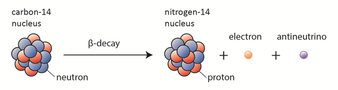
Carbon-14 dating, known also as radiocarbon dating, is a method for determining the age of an object containing organic material by using the properties of radionuclide carbon-14. Radioactive carbon-14 has a half-life of 5730 years and undergoes β− decay, where the neutron is converted into a proton, an electron, and an electron antineutrino:

beta decay - carbon-14 dating
Beta decay of C-14 nucleus.

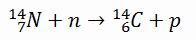
In spite of this short half-life compared to the age of the earth, carbon-14 is a naturally occurring isotope. Its presence can be explained by the following simple observation. Our atmosphere contains many gases, including nitrogen-14. Besides, the atmosphere is constantly bombarded with high energy cosmic rays, consisting of protons, heavier nuclei, or gamma rays. These cosmic rays interact with nuclei in the atmosphere, and produce also high-energy neutrons. These neutrons produced in these collisions can be absorbed by nitrogen-14 to produce an isotope of carbon-14:

carbon-14 dating - formation

Carbon-14 can also be produced in the atmosphere by other neutron reactions, including in particular 13C(n,γ)14C and 17O(n,α)14C. As a result, carbon-14 is continuously formed in the upper atmosphere by the interaction of cosmic rays with atmospheric nitrogen. On average just one out of every 1.3 x 10¹² carbon atoms in the atmosphere is a radioactive carbon-14 atom.

The resulting carbon-14 combines with atmospheric oxygen to form radioactive carbon dioxide, which is incorporated into plants by photosynthesis. Consequently, all biological systems as  plants, animals and humans contain a certain level of radioactive carbon-14. As long as the biological system is alive the level is constant due to constant intake of all isotopes of carbon. When the biological system dies, it stops exchanging carbon with its environment, and from that point onwards the amount of carbon-14 it contains begins to decrease as the carbon-14 undergoes radioactive decay. On the other hand, the amount of stable carbon-12 remains unchanged. As a result, the relative concentration of these two isotopes in any organism changes after its death. The method enables datings to be made up to about 20,000 years ago with an accuracy of about ±100 years.

The technique of carbon dating was suggested initially by Willard Libby and his colleagues in 1949. In 1960, Willard Libby was awarded the Nobel Prize in chemistry for this work.

Age of the Earth – Uranium-lead Dating

The age of the Earth is about 4.54 billion years. This dating is based on evidence from radiometric age-dating of meteorite material and is consistent with the radiometric ages of the oldest-known terrestrial and lunar samples.

One of the oldest radiometric dating methods is uranium-lead dating. The age of the earth’s crust can be estimated from the ratio between the amounts of uranium-238 and lead-206 found in geological specimens. The long half-life of the isotope uranium-238 (4.51×10⁹ years) makes it well-suited for use in estimating the age of the earliest igneous rocks and for other types of radiometric dating, including uranium–thorium dating and uranium–uranium dating.

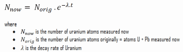
Uranium-lead dating is based on the measurement of the first and the last member of the uranium series, which is one of three classical radioactive series beginning with naturally occurring uranium-238. This radioactive decay chain consists of unstable heavy atomic nuclei that decay through a sequence of alpha and beta decays until a stable nucleus is achieved. In case of uranium series, the stable nucleus is lead-206. The assumption made is that all the lead-206 nuclei found in the specimen today were originally uranium-238 nuclei.  That means at the crust’s formation the specimen contained no lead-206 nuclei. If no other lead isotopes are found in the specimen, this is a reasonable assumption. Under this condition, the age of the sample can be calculated by assuming exponential decay of uranium-238. That is:

uranium-lead method - age of the Earth

Uranium-lead dating method is usually performed on the mineral zircon. Zircons from Jack Hills in Western Australia, have yielded U-Pb ages up to 4.404 billion years, interpreted to be the age of crystallization, making them the oldest minerals so far dated on Earth.

References:

Radiation Protection:

  1. Knoll, Glenn F., Radiation Detection and Measurement 4th Edition, Wiley, 8/2010. ISBN-13: 978-0470131480.
  2. Stabin, Michael G., Radiation Protection and Dosimetry: An Introduction to Health Physics, Springer, 10/2010. ISBN-13: 978-1441923912.
  3. Martin, James E., Physics for Radiation Protection 3rd Edition, Wiley-VCH, 4/2013. ISBN-13: 978-3527411764.
  4. U.S.NRC, NUCLEAR REACTOR CONCEPTS
  5. U.S. Department of Energy, Nuclear Physics and Reactor Theory. DOE Fundamentals Handbook, Volume 1 and 2. January 1993.

Nuclear and Reactor Physics:

  1. J. R. Lamarsh, Introduction to Nuclear Reactor Theory, 2nd ed., Addison-Wesley, Reading, MA (1983).
  2. J. R. Lamarsh, A. J. Baratta, Introduction to Nuclear Engineering, 3d ed., Prentice-Hall, 2001, ISBN: 0-201-82498-1.
  3. W. M. Stacey, Nuclear Reactor Physics, John Wiley & Sons, 2001, ISBN: 0- 471-39127-1.
  4. Glasstone, Sesonske. Nuclear Reactor Engineering: Reactor Systems Engineering, Springer; 4th edition, 1994, ISBN: 978-0412985317
  5. W.S.C. Williams. Nuclear and Particle Physics. Clarendon Press; 1 edition, 1991, ISBN: 978-0198520467
  6. G.R.Keepin. Physics of Nuclear Kinetics. Addison-Wesley Pub. Co; 1st edition, 1965
  7. Robert Reed Burn, Introduction to Nuclear Reactor Operation, 1988.
  8. U.S. Department of Energy, Nuclear Physics and Reactor Theory. DOE Fundamentals Handbook, Volume 1 and 2. January 1993.
  9. Paul Reuss, Neutron Physics. EDP Sciences, 2008. ISBN: 978-2759800414.

See also:

Radioactive Decay

We hope, this article, Radiometric Dating – Radioactive Dating, helps you. If so, give us a like in the sidebar. Main purpose of this website is to help the public to learn some interesting and important information about radiation and dosimeters.

What is Carbon-14 Dating – Radiocarbon Dating – Definition

Carbon-14 dating, known also as radiocarbon dating, is a method for determining the age of an object containing organic material by using the properties of radionuclide carbon-14. Radiation Dosimetry

Carbon-14 dating, known also as radiocarbon dating, is a method for determining the age of an object containing organic material by using the properties of radionuclide carbon-14. Radioactive carbon-14 has a half-life of 5730 years and undergoes β− decay, where the neutron is converted into a proton, an electron, and an electron antineutrino:

beta decay - carbon-14 dating
Beta decay of C-14 nucleus.

In spite of this short half-life compared to the age of the earth, carbon-14 is a naturally occurring isotope. Its presence can be explained by the following simple observation. Our atmosphere contains many gases, including nitrogen-14. Besides, the atmosphere is constantly bombarded with high energy cosmic rays, consisting of protons, heavier nuclei, or gamma rays. These cosmic rays interact with nuclei in the atmosphere, and produce also high-energy neutrons. These neutrons produced in these collisions can be absorbed by nitrogen-14 to produce an isotope of carbon-14:

carbon-14 dating - formation

Carbon-14 can also be produced in the atmosphere by other neutron reactions, including in particular 13C(n,γ)14C and 17O(n,α)14C. As a result, carbon-14 is continuously formed in the upper atmosphere by the interaction of cosmic rays with atmospheric nitrogen. On average just one out of every 1.3 x 10¹² carbon atoms in the atmosphere is a radioactive carbon-14 atom.

The resulting carbon-14 combines with atmospheric oxygen to form radioactive carbon dioxide, which is incorporated into plants by photosynthesis. Consequently, all biological systems as  plants, animals and humans contain a certain level of radioactive carbon-14. As long as the biological system is alive the level is constant due to constant intake of all isotopes of carbon. When the biological system dies, it stops exchanging carbon with its environment, and from that point onwards the amount of carbon-14 it contains begins to decrease as the carbon-14 undergoes radioactive decay. On the other hand, the amount of stable carbon-12 remains unchanged. As a result, the relative concentration of these two isotopes in any organism changes after its death. The method enables datings to be made up to about 20,000 years ago with an accuracy of about ±100 years.

The technique of carbon dating was suggested initially by Willard Libby and his colleagues in 1949. In 1960, Willard Libby was awarded the Nobel Prize in chemistry for this work.

References:

Radiation Protection:

  1. Knoll, Glenn F., Radiation Detection and Measurement 4th Edition, Wiley, 8/2010. ISBN-13: 978-0470131480.
  2. Stabin, Michael G., Radiation Protection and Dosimetry: An Introduction to Health Physics, Springer, 10/2010. ISBN-13: 978-1441923912.
  3. Martin, James E., Physics for Radiation Protection 3rd Edition, Wiley-VCH, 4/2013. ISBN-13: 978-3527411764.
  4. U.S.NRC, NUCLEAR REACTOR CONCEPTS
  5. U.S. Department of Energy, Nuclear Physics and Reactor Theory. DOE Fundamentals Handbook, Volume 1 and 2. January 1993.

Nuclear and Reactor Physics:

  1. J. R. Lamarsh, Introduction to Nuclear Reactor Theory, 2nd ed., Addison-Wesley, Reading, MA (1983).
  2. J. R. Lamarsh, A. J. Baratta, Introduction to Nuclear Engineering, 3d ed., Prentice-Hall, 2001, ISBN: 0-201-82498-1.
  3. W. M. Stacey, Nuclear Reactor Physics, John Wiley & Sons, 2001, ISBN: 0- 471-39127-1.
  4. Glasstone, Sesonske. Nuclear Reactor Engineering: Reactor Systems Engineering, Springer; 4th edition, 1994, ISBN: 978-0412985317
  5. W.S.C. Williams. Nuclear and Particle Physics. Clarendon Press; 1 edition, 1991, ISBN: 978-0198520467
  6. G.R.Keepin. Physics of Nuclear Kinetics. Addison-Wesley Pub. Co; 1st edition, 1965
  7. Robert Reed Burn, Introduction to Nuclear Reactor Operation, 1988.
  8. U.S. Department of Energy, Nuclear Physics and Reactor Theory. DOE Fundamentals Handbook, Volume 1 and 2. January 1993.
  9. Paul Reuss, Neutron Physics. EDP Sciences, 2008. ISBN: 978-2759800414.

See also:

Radiometric Dating

We hope, this article, Carbon-14 Dating – Radiocarbon Dating, helps you. If so, give us a like in the sidebar. Main purpose of this website is to help the public to learn some interesting and important information about radiation and dosimeters.

What is Age of the Earth – How old is the Earth – Definition

The age of the Earth is about 4.54 billion years. The oldest zircons from Jack Hills in Western Australia have yielded U-Pb ages up to 4.404 billion years, interpreted to be the age of crystallization. Radiation Dosimetry

The age of the Earth is about 4.54 billion years. This dating is based on evidence from radiometric age-dating of meteorite material and is consistent with the radiometric ages of the oldest-known terrestrial and lunar samples.

One of the oldest radiometric dating methods is uranium-lead dating. The age of the earth’s crust can be estimated from the ratio between the amounts of uranium-238 and lead-206 found in geological specimens. The long half-life of the isotope uranium-238 (4.51×10⁹ years) makes it well-suited for use in estimating the age of the earliest igneous rocks and for other types of radiometric dating, including uranium–thorium dating and uranium–uranium dating.

Uranium-lead dating is based on the measurement of the first and the last member of the uranium series, which is one of three classical radioactive series beginning with naturally occurring uranium-238. This radioactive decay chain consists of unstable heavy atomic nuclei that decay through a sequence of alpha and beta decays until a stable nucleus is achieved. In case of uranium series, the stable nucleus is lead-206. The assumption made is that all the lead-206 nuclei found in the specimen today were originally uranium-238 nuclei.  That means at the crust’s formation the specimen contained no lead-206 nuclei. If no other lead isotopes are found in the specimen, this is a reasonable assumption. Under this condition, the age of the sample can be calculated by assuming exponential decay of uranium-238. That is:

uranium-lead method - age of the Earth

Uranium-lead dating method is usually performed on the mineral zircon. Zircons from Jack Hills in Western Australia, have yielded U-Pb ages up to 4.404 billion years, interpreted to be the age of crystallization, making them the oldest minerals so far dated on Earth.

References:

Radiation Protection:

  1. Knoll, Glenn F., Radiation Detection and Measurement 4th Edition, Wiley, 8/2010. ISBN-13: 978-0470131480.
  2. Stabin, Michael G., Radiation Protection and Dosimetry: An Introduction to Health Physics, Springer, 10/2010. ISBN-13: 978-1441923912.
  3. Martin, James E., Physics for Radiation Protection 3rd Edition, Wiley-VCH, 4/2013. ISBN-13: 978-3527411764.
  4. U.S.NRC, NUCLEAR REACTOR CONCEPTS
  5. U.S. Department of Energy, Nuclear Physics and Reactor Theory. DOE Fundamentals Handbook, Volume 1 and 2. January 1993.

Nuclear and Reactor Physics:

  1. J. R. Lamarsh, Introduction to Nuclear Reactor Theory, 2nd ed., Addison-Wesley, Reading, MA (1983).
  2. J. R. Lamarsh, A. J. Baratta, Introduction to Nuclear Engineering, 3d ed., Prentice-Hall, 2001, ISBN: 0-201-82498-1.
  3. W. M. Stacey, Nuclear Reactor Physics, John Wiley & Sons, 2001, ISBN: 0- 471-39127-1.
  4. Glasstone, Sesonske. Nuclear Reactor Engineering: Reactor Systems Engineering, Springer; 4th edition, 1994, ISBN: 978-0412985317
  5. W.S.C. Williams. Nuclear and Particle Physics. Clarendon Press; 1 edition, 1991, ISBN: 978-0198520467
  6. G.R.Keepin. Physics of Nuclear Kinetics. Addison-Wesley Pub. Co; 1st edition, 1965
  7. Robert Reed Burn, Introduction to Nuclear Reactor Operation, 1988.
  8. U.S. Department of Energy, Nuclear Physics and Reactor Theory. DOE Fundamentals Handbook, Volume 1 and 2. January 1993.
  9. Paul Reuss, Neutron Physics. EDP Sciences, 2008. ISBN: 978-2759800414.

See also:

Radiometric Dating

We hope, this article, Age of the Earth – How old is the Earth, helps you. If so, give us a like in the sidebar. Main purpose of this website is to help the public to learn some interesting and important information about radiation and dosimeters.