The neutron life cycle quantifies the most important neutron-physical processes that occur in the nuclear reactor. These processes of neutron life cycle are quantified by their factors. Radiation Dosimetry
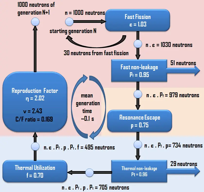
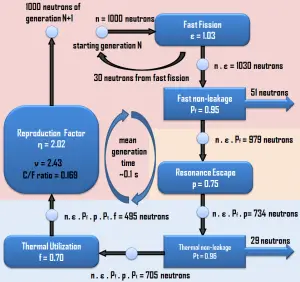
The neutron life cycle quantifies the most important neutron-physical processes that occur in the nuclear reactor. In the reactor, neutrons are produced as fast neutrons and most of them are absorbed after thermalization. There are six processes (factors) that describe the inherent multiplication ability of the system. Four of them are completely independent of the size and shape of the reactor and these are:
Fast Fission. The fast fission process is in the multiplication factor characterized by the fast fission factor, ε, which increases the fast neutron population in one neutron generation. The fast fission factor is defined as the ratio of the fast neutrons produced by fissions at all energies to the number of fast neutrons produced in thermal fission.
Resonance Escape. The resonance escape probability, symbolized by p, is the probability that a neutron will be slowed to thermal energy and will escape resonance capture. This probability is defined as the ratio of the number of neutrons that reach thermal energies to the number of fast neutrons that start to slow down.
Thermal Utilization. The thermal utilisation factor, f, is the fraction of the thermal neutrons that are absorbed in the nuclear fuel, in all isotopes of the nuclear fuel. It describes how effectively (how well are utilized) are thermal neutrons absorbed in the fuel. The value of the thermal utilization factor is given by the ratio of the number of thermal neutrons absorbed in the fuel (all nuclides) to the number of thermal neutrons absorbed in all the material that makes up the core.
Reproduction. The number of neutrons created in the new generation is determined by the neutron reproduction factor. The reproduction factor, η, is defined as the ratio of the number of fast neutrons produced by thermal fission to the numberof thermal neutrons absorbed in the fuel.
These processes of neutron life cycle, quantified by their factors, constitute the infinite multiplication factor (k∞), which may be expressed mathematically in terms of these factors by following equation, usually known as the four factor formula:
k∞ = η.ε.p.f
The effective multiplication factor (keff) may be expressed mathematically in terms of the infinite multiplication factor (k∞) and two additional factors which account for neutron leakage during neutron thermalisation (fast non-leakage probability) and neutron leakage during neutron diffusion (thermal non-leakage probability) by following equation, usually known as the six factor formula:
keff = k∞ . Pf . Pt
Fast Non-leakage Probability. During the slowing down process, some of the neutrons leak out of the boundaries of the reactor core before they become thermalized. This process and its impact on the effective multiplying factor is characterized by the fast non-leakage factor, Pf, which is defined as the ratio of the number of fast neutrons that do not leak from the reactor core during the slowing down process to the number of fast neutrons produced by fissions at all energies.
Thermal Non-leakage Probability. During the neutron diffusion, some of the neutrons leak out of the boundaries of the reactor core before they are absorbed. This process and its impact on the effective multiplying factor is characterized by the thermal non-leakage factor, Pt, which is defined as the ratio of the number of thermal neutrons that do not leak from the reactor core during the neutron diffusion process to the number of neutrons that reach thermal energies.
In reactor physics, keff is the most significant parameter with regard to reactor control. At any specific power level or condition of the reactor, keff is kept as near to the value of 1.0 as possible. At this point in operation, the neutron balance is kept to exactly one neutron completing the life cycle for each original neutron absorbed in the fuel.
Neutron Life Cycle in Fast Reactors
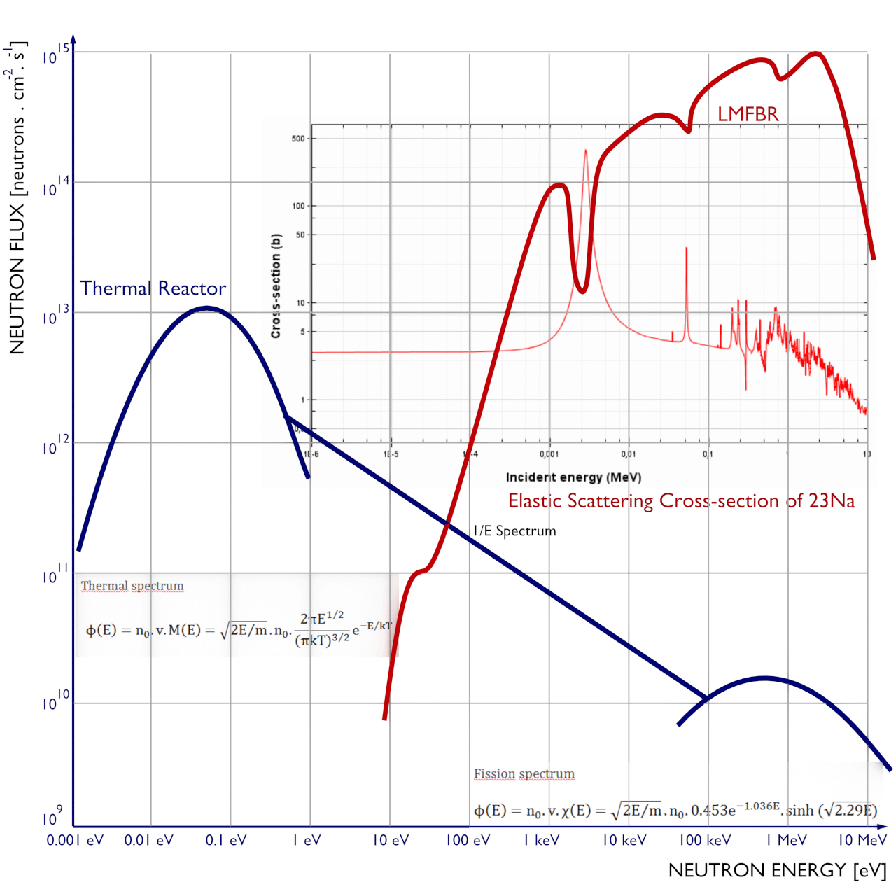
The spectrum of neutron energies produced by fission vary significantly with certain reactor design. thermal vs. fast reactor neutron spectrum
The method of calculations of multiplication factors has been developed in the early years of nuclear energy and is only applicable to thermal reactors, where the bulk of fission reactions occurs at thermal energies. This method well puts into the context all the processes, that are associated with the thermal reactors (e.g. the neutron thermalisation, the neutron diffusion or the fast fission), because the most important neutron-physical processes occur in energy regions that can be clearly separated from each other. In short, the calculation of multiplication factor gives a good insight in the processes that occur in each thermal multiplying system.
For fast reactors, in which the fission are caused by neutrons with a very broad energy distribution, such an analysis is inappropriate. The neutron flux in fast reactors have to be divided into many energy groups. Moreover, in fast reactors, the neutron thermalisation is undesirable process and therefore the four factor formula does not really make any sense. The resonance escape probability is not significant because very few neutrons exist at energies where resonance absorption is significant. The thermal non-leakage probability does not exist because the reactor is designed to avoid the thermalization of neutrons.
References:
Nuclear and Reactor Physics:
J. R. Lamarsh, Introduction to Nuclear Reactor Theory, 2nd ed., Addison-Wesley, Reading, MA (1983).
J. R. Lamarsh, A. J. Baratta, Introduction to Nuclear Engineering, 3d ed., Prentice-Hall, 2001, ISBN: 0-201-82498-1.
W. M. Stacey, Nuclear Reactor Physics, John Wiley & Sons, 2001, ISBN: 0- 471-39127-1.
Robert Reed Burn, Introduction to Nuclear Reactor Operation, 1988.
U.S. Department of Energy, Nuclear Physics and Reactor Theory. DOE Fundamentals Handbook, Volume 1 and 2. January 1993.
Advanced Reactor Physics:
K. O. Ott, W. A. Bezella, Introductory Nuclear Reactor Statics, American Nuclear Society, Revised edition (1989), 1989, ISBN: 0-894-48033-2.
K. O. Ott, R. J. Neuhold, Introductory Nuclear Reactor Dynamics, American Nuclear Society, 1985, ISBN: 0-894-48029-4.
D. L. Hetrick, Dynamics of Nuclear Reactors, American Nuclear Society, 1993, ISBN: 0-894-48453-2.
E. E. Lewis, W. F. Miller, Computational Methods of Neutron Transport, American Nuclear Society, 1993, ISBN: 0-894-48452-4.
See also:
See also:
Chain Reaction
See also:
We hope, this article, Neutron Life Cycle, helps you. If so, give us a like in the sidebar. Main purpose of this website is to help the public to learn some interesting and important information about radiation and dosimeters.
If you think you are reaching out to all of your online prospects, think again! Here you will find all the information you need to advertise on Nuclear Power. Advertise Here
If you think you are reaching out to all of your online prospects, think again! Here you will find all the information you need to advertise on Nuclear Power.
The main purpose of this website is to promote the awareness and understanding with regard to the application of nuclear science and technology. This website contains more than 1500 articles about nuclear engineering, reactor physics, hydraulics, thermodynamics or radiation protection. It was a really hard work and the work will continue. Moreover, we would like to offer multilinguality of our website in next few years. But it is a very extensive work and funds are minimal. Over the last 12 months, the Nuclear Power website has attracted:
more than million unique visitors
corresponding to about two millions potential banner/page views.
The demographic composition of our visitors is obvious:
about 85.2% of visitors are from organic search
about 50% of visitors are 18-24 years old (students)
about 30% of visitors are 25-34 years old (young professionals)
about 30% of visitors are from the United States
about 25% of visitors are from India
the UK, Canada, Australia follows
Simply, most of our visitors are young technicians, physicists, engineers, who discover the world of energy and who need new connections in powery industry. You can sponsor one or more of the pages on this site by displaying your banner ad at the top of the page or in the sidebar, linking directly to your corporate website (or any other URL that you want).
These prices are in costs per mille (CPM), that means $X per 1000 displays. We accept payments via Wire transfer or PayPal. Banner advertising on nuclear-power.com is a very cost effective way to showcase your services to the rest of power industry. We are selective with partners, choosing only those companies/services that meet our requirements and requirements of our partners.
Contact Us
If you want know more, please do not hesitate to contact us via e-mail:
info@www.nuclear-power.com
We hope, this article, Advertise Here, helps you. If so, give us a like in the sidebar. Main purpose of this website is to help the public to learn some interesting and important information about radiation and dosimeters.
Detailed knowledge about detection of radiation is very important in many branches of engineering, including radiation protection. Radiation Detection – Detection of Ionization Radiation
Source of gamma radiation. The danger of ionizing radiation lies in the fact that the radiation is invisible and not directly detectable by human senses.
The danger of ionizing radiation lies in the fact that the radiation is invisible and not directly detectable by human senses. People can neither see nor feel radiation, yet it deposits energy to the molecules of material. The energy is transferred in small quantities for each interaction between the radiation and a molecule and there are usually many types of interactions. Therefore, the only way you can detect and measure radiation is to use instruments (detectors of ionizing radiation).
Detailed knowledge about detection of radiation is very important in many branches of engineering, including radiation protection. Most modern nuclear or particle experiments use a variety of sophisticated detectors for measuring and detection of sub-atomic particles. In order to be detected, a particle must leave some trace of its presence in a detector. Particles mostly deposit energy along its path. Knowledge of this interaction, how different particles deposit energy in the matter and how much energy particles deposit, is fundamental for our understanding of many problems. This chapter will give you a basic understanding of how these detectors work and some of their limitations.
What is Ionization
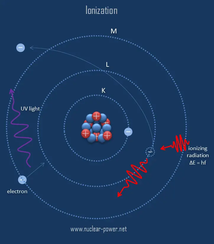
Ionization is the process in which an atom or a molecule gains or loses electrons to form charged ion. Ionization can result from the loss of an electron after collisions with energetic subatomic particles, collisions with other atoms, molecules and ions, or through the interaction with electromagnetic radiation. In general, ionizing radiation is any radiation (particles or electromagnetic waves) that carries enough energy to knock electrons from atoms or molecules, thereby ionizing them. For ionizing radiation, the kinetic energy of particles (photons, electrons, etc.) is sufficient and the particle can ionize (to form ion by losing electrons) target atoms to form ions.
The boundary between ionizing and non-ionizing radiation is not sharply defined, since different molecules and atoms ionize at different energies. Gamma rays, X-rays, and the higher ultraviolet part of the spectrum are ionizing, whereas the lower ultraviolet, visible light (including laser light), infrared, microwaves, and radio waves are considered non-ionizing radiation.
Basic Principles of Detectors
Detectors of ionizing radiation consist of two parts that are usually connected. The first part consists of a sensitive material, consisting of a compound that experiences changes when exposed to radiation. The other component is a device that converts these changes into measurable signals.
In their basic principles of operation, most detectors of ionizing radiation follow similar characteristics. Detectors of ionizing radiation consist of two parts that are usually connected. The first part consists of a sensitive material, consisting of a compound that experiences changes when exposed to radiation. The other component is a device that converts these changes into measurable signals. All detectors require that radiation must deposit some of its energy in sensitive material that forms part of the instrument. The radiation enters the detector, interacts with atoms of the detector material and deposits some energy to sensitive material. Each event may generate a signal, which can be a pulse, hole, light signal, ion pairs in a gas, and many others. The main task is to generate sufficient signal, amplify it and to record it.
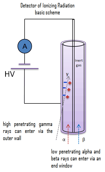
Let assume gaseous ionization detectors. Basic gaseous ionization detector consists of a chamber that is filled with a suitable medium (air or a special fill gas) that can be easily ionized. As a general rule, the center wire is the positive electrode (anode) and the outer cylinder is the negative electrode (cathode), so that (negative) electrons are attracted to the center wire and positive ions are attracted to the outer cylinder. The anode is at a positive voltage with respect to the detector wall. As ionizing radiation enters the gas between the electrodes, a finite number of ion-pairs are formed. Under the influence of the electric field, the positive ions will move toward the negatively charged electrode (outer cylinder), and the negative ions (electrons) will migrate toward the positive electrode (central wire). The collection of these ions will produce a charge on the electrodes and an electrical pulse across the detection circuit. However it is a small signal, this signal can be amplified, and then recorded using standard electronics.
Classification of Radiation Detectors – Purpose
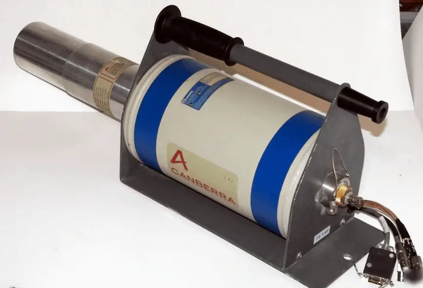
HPGe detector with LN2 cryostat Source: canberra.com
Generally, detectors of ionizing radiation may be classified according to their purpose. Their purpose is closely related with the generated signal in the detector. There are three main types of detectors, which record different types of signals.
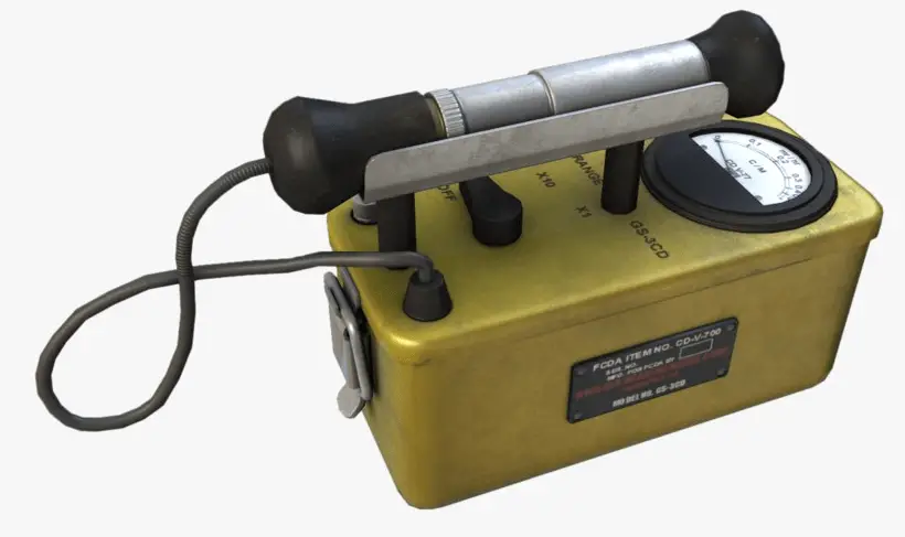
Counter. The activity or intensity of radiation is measured in counts per second (cps). The best known counter is the Geiger-Müller counter. In radiation counters the generated signal from the incident radiation is created by the counting of the number of interactions occurring at the sensitive volume of the detector.
Radiation Spectrometer. Spectrometers are devices designed to measure the spectral power distribution of a source. The incident radiation generates a signal that allows to determine the energy of the incident particle.
Dosimeter. A radiation dosimeter is a device that measures exposure to ionizing radiation. Dosimeters usually record a dose, which is the absorbed radiation energy measured in grays (Gy) of the equivalent dose measured in sieverts (Sv) A personal dosimeter is dosimeter, that is worn at the surface of the body by the person being monitored, and it records of the radiation dose received.
Classification of Radiation Detectors – Radiation Type
Principles and methods for detection of ionizing radiation are dependent on many factors. Type of radiation measured and detected is a key factor, and different types of detectors in different physical states (solid, liquid or gas) are used to measure selective types of ionizing radiation. It is important to distinguish between alpha or beta particles, X-rays or γ-rays and neutrons. Since each radiation interacts differently with matter, we cannot use, for example, neutron detector to detect beta radiation. Sometimes, detectors can detect various types of radiation, for example, neutron detectors are also able to detect gamma radiation. Usually, this feature is not desirable, and detectors must compensate the undesirable component of radiation.
In order to describe the classification of radiation detectors by the type of radiation, we have to understand the interactions of radiation with matter. Each type particle interacts in a different way, therefore we must describe interaction of particles (radiation as a flow of these particles) separately. For example charged particles with high energies can directly ionize atoms. On the other hand electrically neutral particles interacts only indirectly, but can also transfer some or all of their energies to the matter. This is the key feature of the categorization of radiation detectors. Simply, we cannot use a gamma rays detector to detect alpha radiation. Detectors can be categorized into two general types as follows:
Robert Reed Burn, Introduction to Nuclear Reactor Operation, 1988.
U.S. Department of Energy, Nuclear Physics and Reactor Theory. DOE Fundamentals Handbook, Volume 1 and 2. January 1993.
Paul Reuss, Neutron Physics. EDP Sciences, 2008. ISBN: 978-2759800414.
See also:
Engineering
We hope, this article, Radiation Detection, helps you. If so, give us a like in the sidebar. Main purpose of this website is to help the public to learn some interesting and important information about radiation and dosimeters.
Detectors of ionizing radiation consist of two parts that are usually connected. The first part consists of a sensitive material, consisting of a compound that experiences changes when exposed to radiation. Radiation Dosimetry
Detector of Ionizing Radiation – Geiger Tube
Various instruments and devices are used for the detection of radiation. Generally, detectors of ionizing radiation may be classified according to their purpose. Its purpose is closely related with the generated signal in the detector. There are three main types of detectors, which record different types of signals.
Counter. The activity or intensity of radiation is measured in counts per second (cps). The best known counter is the Geiger-Müller counter. In radiation counters the generated signal from the incident radiation is created by the counting of the number of interactions occurring at the sensitive volume of the detector.
Radiation Spectrometer. Spectrometers are devices designed to measure the spectral power distribution of a source. The incident radiation generates a signal that allows to determine the energy of the incident particle.
Dosimeter. A radiation dosimeter is a device that measures exposure to ionizing radiation. Dosimeters usually record a dose, which is the absorbed radiation energy measured in grays (Gy) of the equivalent dose measured in sieverts (Sv) A personal dosimeter is dosimeter, that is worn at the surface of the body by the person being monitored, and it records of the radiation dose received.
All these types of equipment require that the radiations result in observable changes in a compound (whether gas, liquid or solid). In their basic principles of operation, most detectors of ionizing radiation follow similar characteristics. Detectors of ionizing radiation consist of two parts that are usually connected. The first part consists of a sensitive material, consisting of a compound that experiences changes when exposed to radiation. The other component is a device that converts these changes into measurable signals. All detectors require that radiation must deposit some of its energy in sensitive material that forms part of the instrument. The radiation enters the detector, interacts with atoms of the detector material and deposits some energy to sensitive material. Each event may generate a signal, which can be a pulse, hole, light signal, ion pairs in a gas, and many others. The main task is to generate sufficient signal, amplify it and to record it.
In this section we describe a number of different types of radiation measuring instruments. We focus on the first part, which consists of a sensitive material. Detectors may be categorized according to sensitive materials and methods that can be utilized to make a measurement:
Gaseous Ionization Detectors
Scintillation Detectors
Semiconductor Detectors
Film Badges
TLD
Basic Principles of Detectors
Detectors of ionizing radiation consist of two parts that are usually connected. The first part consists of a sensitive material, consisting of a compound that experiences changes when exposed to radiation. The other component is a device that converts these changes into measurable signals.
In their basic principles of operation, most detectors of ionizing radiation follow similar characteristics. Detectors of ionizing radiation consist of two parts that are usually connected. The first part consists of a sensitive material, consisting of a compound that experiences changes when exposed to radiation. The other component is a device that converts these changes into measurable signals. All detectors require that radiation must deposit some of its energy in sensitive material that forms part of the instrument. The radiation enters the detector, interacts with atoms of the detector material and deposits some energy to sensitive material. Each event may generate a signal, which can be a pulse, hole, light signal, ion pairs in a gas, and many others. The main task is to generate sufficient signal, amplify it and to record it.
Let assume gaseous ionization detectors. Basic gaseous ionization detector consists of a chamber that is filled with a suitable medium (air or a special fill gas) that can be easily ionized. As a general rule, the center wire is the positive electrode (anode) and the outer cylinder is the negative electrode (cathode), so that (negative) electrons are attracted to the center wire and positive ions are attracted to the outer cylinder. The anode is at a positive voltage with respect to the detector wall. As ionizing radiation enters the gas between the electrodes, a finite number of ion-pairs are formed. Under the influence of the electric field, the positive ions will move toward the negatively charged electrode (outer cylinder), and the negative ions (electrons) will migrate toward the positive electrode (central wire). The collection of these ions will produce a charge on the electrodes and an electrical pulse across the detection circuit. However it is a small signal, this signal can be amplified, and then recorded using standard electronics.
Signal-to-Noise Ratio
Signal-to-noise ratio, SNR, is a measure used in science and engineering that compares the electrical output signal to the electrical noise generated in the cable run or in the instrumentation.
Discriminator
Discrimination in radiation detection circuits refers to the process of distinguishing between different types of radiation on the basis of pulse height. A discriminator circuit selects the minimum or maximum pulse height that is to be counted. For example a discriminator circuit is used in the neutron detector to distinguish the large pulses from alpha ionisation (neutron absorption in the detector produces alpha particles) from the much smaller ones that would be caused by gamma rays.
Special Reference: U.S. Department of Energy, Instrumantation and Control. DOE Fundamentals Handbook, Volume 2 of 2. June 1992.
Robert Reed Burn, Introduction to Nuclear Reactor Operation, 1988.
U.S. Department of Energy, Nuclear Physics and Reactor Theory. DOE Fundamentals Handbook, Volume 1 and 2. January 1993.
Paul Reuss, Neutron Physics. EDP Sciences, 2008. ISBN: 978-2759800414.
See also:
Detection
We hope, this article, Detector of Ionization Radiation, helps you. If so, give us a like in the sidebar. Main purpose of this website is to help the public to learn some interesting and important information about radiation and dosimeters.
Gaseous ionization detectors are radiation detection instruments used in radiation protection applications to measure ionizing radiation, and also in particle physics to detect the presence of ionizing particles. Radiation Dosimetry
Detector of Ionizing Radiation – Geiger Tube
Gaseous ionization detectors are radiation detection instruments used in radiation protection applications to measure ionizing radiation, and also in particle physics to detect the presence of ionizing particles. These detectors are designed to measure the ionization produced when an incident particle traverses some medium, they are based on the ionising effect of radiation. For ionizing radiation to occur, the kinetic energy of particles (photons, electrons, etc.) of ionizing radiation is sufficient and the particle can ionize (to form ion by losing electrons) target atoms to form ions. Simply ionizing radiation can knock electrons from an atom.
The basic gaseous ionization detector consists of a chamber that is filled with a suitable medium (air or a special fill gas) that can be easily ionized. Most widely used types of these detectors are based on the effects produced when a charged particle passes through a gas. The operating medium:
should be chemically stable (or inert) so that the moving ionization electrons are not easily captured by the molecules of that medium
should have a low value of ionization potential (I) in order to maximize the amount of ionization produced per energy deposited by any incident particle.
should not be very sensitive to radiation damage so that its response to incident particles does not change markedly with use.
Typical gases used in detectors are argon and helium, although boron-triflouride (BF3) is utilized when the detector is to be used to measure neutrons. Gaseous ionization detectors are widely used in nuclear power plants, for the most part, to measure alpha and beta particles, neutrons, and gamma rays. The detectors operate in the ionization, proportional, and Geiger-Mueller regions with an arrangement most sensitive to the type of radiation being measured. Neutron detectors utilize ionization chambers or proportional counters of appropriate design. Compensated ion chambers, BF3 counters, fission counters, and proton recoil counters are examples of neutron detectors.
Basic Principle of Gaseous Ionization Detectors
Detectors of ionizing radiation consist of two parts that are usually connected. The first part consists of a sensitive material, consisting of a compound that experiences changes when exposed to radiation. The other component is a device that converts these changes into measurable signals.
The chamber has a cathode and an anode that are held at some large relative voltage, and the device is characterized by a capacitance that is determined by the geometry of the electrodes. As ionizing radiation enters the gas between the electrodes, a finite number of ion-pairs are formed. The behavior of the resultant ion-pairs is affected by the potential gradient of the electric field within the gas and the type and pressure of the fill gas. Under the influence of the electric field, the positive ions will move toward the negatively charged electrode (outer cylinder), and the negative ions (electrons) will migrate toward the positive electrode (central wire). The electric field in this region keeps the ions from recombining with the electrons. The collection of these ions will produce a charge on the electrodes and an electrical pulse across the detection circuit. In air, the average energy needed to produce an ion is about 34 eV, therefore a 1 MeV radiation completely absorbed in the detector produces about 3 x 10⁴ pair of ions. However it is a small signal, this signal can be considerably amplified using standard electronics.
Operating Regions of Ionizing Detectors – Detector Voltage
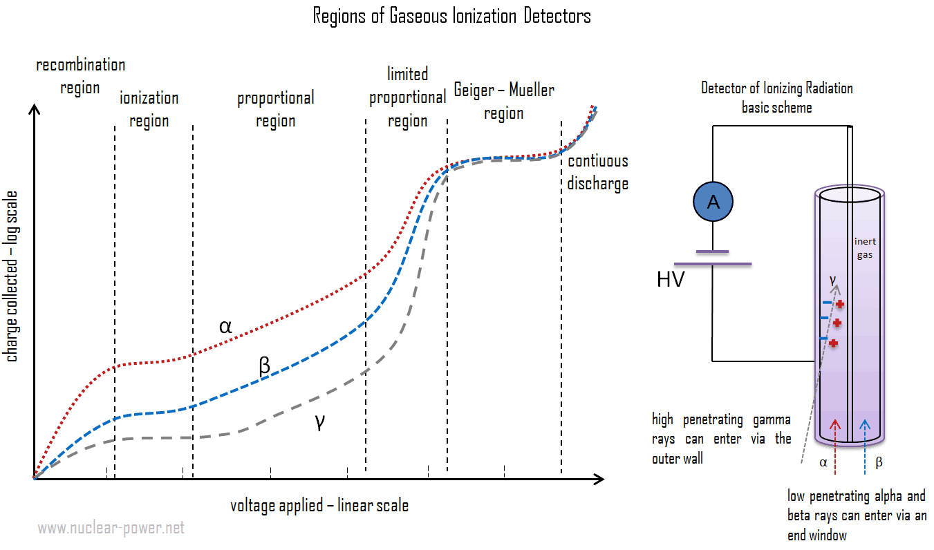
The relationship between the applied voltage and pulse height in a detector is very complex. Pulse height and the number of ion pairs collected are directly related. As was written, the voltages can vary widely depending upon the detector geometry and the gas type and pressure. The figure schematically indicates the different voltage regions for alpha, beta and gamma rays. There are six main practical operating regions, where three (ionization, proportional and Geiger-Mueller region) are useful to detect ionizing radiation. These reqions are shown below. The alpha curve is higher than the beta and gamma curve from recombination region to part of limited proportionality region due to the larger number of ion pairs produced by the initial reaction of the incident radiation.
This diagram shows the number of ion pairs generated in the gas-filled detector, which varies according to the applied voltage for constant incident radiation. The voltages can vary widely depending upon the detector geometry and the gas type and pressure. This figure schematically indicates the different voltage regions for alpha, beta and gamma rays. There are six main practical operating regions, where three (ionization, proportional and Geiger-Mueller region) are useful to detect ionizing radiation. Alpha particles are more ionising than beta particles and than gamma rays, so more current is produced in the ion chamber region by alpha than beta and gamma, but the particles cannot be differentiated. More current is produced in the proportional counting region by alpha particles than beta, but by the nature of proportional counting it is possible to differentiate alpha, beta and gamma pulses. In the Geiger region, there is no differentiation of alpha and beta as any single ionisation event in the gas results in the same current output.
Recombination Region. At low voltage, the electric field is not large enough to accelerate electrons and ions. The electrons and ions can recombine soon after they are produced, and only a small fraction of the produced electrons and ions reach their respective electrodes. As the detector voltage is increased, however, an increasingly large fraction of the ions produced will reach the electrodes. This increase continues until the “saturation” voltage is attained. The range of operating voltage where this occurs is referred to as the recombination region. Detectors are not operated in this region, because neither the number of recombinations nor the number of ion-pairs initially produced can be determined accurately.
Ionization Region. In the ionization region, an increase in voltage does not cause a substantial increase in the number of ion-pairs collected. The number of ion-pairs collected by the electrodes is equal to the number of ion-pairs produced by the incident radiation, and is dependent on the type and energy of the particles or rays in the incident radiation. Therefore, in this region the curve is flat. The voltage must be higher than the point where dissociated ion-pairs can recombine. On the other hand, the voltage is not high enough to produce gas amplification (secondary ionization). Detectors in the ionization region operate at a low electric field strength, selected such that no gas multiplication takes place. Their current is independent of the applied voltage, and they are preferred for high radiation dose rates because they have no “dead time”, a phenomenon which affects the accuracy of the Geiger-Mueller tube at high dose rates.
Proportional Region. In the proportional region, the charge collected increases with a further increase in the detector voltage, while the number of primary ion-pairs remains unchanged. Increasing the voltage, provides the primary electrons with sufficient acceleration and energy so that they can ionize additional atoms of the medium. These secondary ions formed are also accelerated causing an effect known as Townsend avalanches, which creates a single large electrical pulse. Even though there is a large number of secondary ions (about 103 – 105) for each primary event, the chamber is always operated such that the number of secondary ions is proportional to the number of primary events. It is very important, because the primary ionization is dependent on the type and energy of the particles or rays in the intercepted radiation field. The number of ion pairs collected divided by the number of ion pairs produced by the primary ionization provides the gas amplification factor (denoted by A). The gas amplification that occurs in this region can increase the total amount of ionization to a measurable value. The process of charge amplification greatly improves the signal-to-noise ratio of the detector and reduces the subsequent electronic amplification required. When instruments are operated in the proportional region, the voltage must be kept constant. If a voltage remains constant the gas amplification factor also does not change. Proportional counter detection instruments are very sensitive to low levels of radiation. Moreover, proportional counters are capable of particle identification and energy measurement (spectroscopy). Different energies of radiation and different types of radiation can be distinguished by analyzing the pulse height, since they significantly differ in the primary ionization.
Limited Proportional Region. In the limited proportional region, the gas amplification factor does not continue to increase proportionally to the voltage. Additional ionizations and nonlinear effects causes, that there is no proportionality of the output signal to the deposited energy at a given applied voltage. The electric field in the chamber is distorted due to the high positive ion concentration. Free electrons are much lighter than the positive ions, thus, they are drawn toward the positive central electrode much faster than the positive ions are drawn to the chamber wall. The resulting cloud of positive ions near the electrode leads to distortions in gas multiplication. This region is usually avoided as a detection region.
Geiger-Mueller Region. In the Geiger-Mueller region, the voltage and thus the electric field is so strong that secondary avalanches can occur. These avalanches can be triggered and propagated by photons emitted by atoms excited in the original avalanche. Since these photons are not affected by the electric field, they may interact far (e.g. laterally to the axis) from the primary avalanche, the entire Geiger tube is participating in the process. A strong signal (the amplification factor can reach about 1010) is produced by these avalanches with shape and height independently of the primary ionization and the energy of the detected photon. Detectors, which are operated in the Geiger-Mueller region, are capable of detection of gamma rays, and also of all types of charged particles, that can enter the detector. These detectors are known as Geiger counters. Main advantage of these instruments is that they usually do not require any signal amplifiers. Since the positive ions do not move far from the avalanche region, a positively charged ion cloud disturbs the electric field and terminates the avalanche process. In practice the termination of the avalanche is improved by the use of “quenching” techniques. In contrast to proportional counters the energy or even incident radiation particle cannot not be distinguished by Geiger counters, since the output signal is independent of the amount and type of original ionization.
Discharge Region. Finally, at still higher voltages (above Geiger-Mueller region), the electric field generates a continuous discharge of the medium, with the chamber no longer being sensitive to any incident ionization. This region is not used for detection or measurement of ionizing radiation. If the Geiger tube voltage is increased above the end of the plateau the count rate begins to increase rapidly again, until the onset of continuous discharge where the tube cannot detect radiation, and may be damaged.
Types of Detectors of Ionizing Radiation
As a result, there are three basic types of gaseous ionization detectors, which are categorized according to the voltage applied to the detector:
ionization chambers,
proportional counters,
Geiger-Müller tubes.
As with other detectors, ionization chambers can be operated in current or pulse mode. In contrast, proportional counters or Geiger counters are almost always used in pulse mode. Detectors of ionizing radiation can be used both for activity measurements as well as for dose measurement. With knowledge about the energy needed to form an pair of ions – the dose can be obtained.
Robert Reed Burn, Introduction to Nuclear Reactor Operation, 1988.
U.S. Department of Energy, Nuclear Physics and Reactor Theory. DOE Fundamentals Handbook, Volume 1 and 2. January 1993.
Paul Reuss, Neutron Physics. EDP Sciences, 2008. ISBN: 978-2759800414.
See also:
Detection
We hope, this article, Gaseous Ionization Detector – Gas-Filled Detector, helps you. If so, give us a like in the sidebar. Main purpose of this website is to help the public to learn some interesting and important information about radiation and dosimeters.
The ionization chamber, also known as the ion chamber, is electrical device that detects various types of ionizing radiation. The voltage of detector is adjusted so that the conditions correspond to the ionization region. Radiation Dosimetry
The ionization chamber, also known as the ion chamber, is electrical device that detects various types of ionizing radiation. The voltage of detector is adjusted so that the conditions correspond to the ionization region. The voltage is not high enough to produce gas amplification (secondary ionization). Detectors in the ionization region operate at a low electric field strength, selected such that no gas multiplication takes place. The charge collected (output signal) is independent of the applied voltage and for single minimum-ionizing particles tends to be quite small and usually require special low-noise amplifiers for attaining efficient operating performance. Ionization chambers are preferred for high radiation dose rates because they have no “dead time”, a phenomenon which affects the accuracy of the Geiger-Mueller tube at high dose rates. This is due to the fact, there is no inherent amplification of signal in the operating medium and therefore these types of counters do not require much time to recover from large currents. In addition, because there is no amplification, they provide excellent energy resolution, which is limited primarily by electronic noise.
Ionization chambers can be operated in current or pulse mode. In contrast, proportional counters or Geiger counters are almost always used in pulse mode. Detectors of ionizing radiation can be used both for activity measurements as well as for dose measurement. With knowledge about the energy needed to form an pair of ions – the dose can be obtained.
Ionization Region
This diagram shows the number of ion pairs generated in the gas-filled detector, which varies according to the applied voltage for constant incident radiation. The voltages can vary widely depending upon the detector geometry and the gas type and pressure. This figure schematically indicates the different voltage regions for alpha, beta and gamma rays. There are six main practical operating regions, where three (ionization, proportional and Geiger-Mueller region) are useful to detect ionizing radiation. Alpha particles are more ionising than beta particles and than gamma rays, so more current is produced in the ion chamber region by alpha than beta and gamma, but the particles cannot be differentiated. More current is produced in the proportional counting region by alpha particles than beta, but by the nature of proportional counting it is possible to differentiate alpha, beta and gamma pulses. In the Geiger region, there is no differentiation of alpha and beta as any single ionisation event in the gas results in the same current output.
In the ionization region, an increase in voltage does not cause a substantial increase in the number of ion-pairs collected. The number of ion-pairs collected by the electrodes is equal to the number of ion-pairs produced by the incident radiation, and is dependent on the type and energy of the particles or rays in the incident radiation. Therefore, in this region the curve is flat. The voltage must be higher than the point where dissociated ion-pairs can recombine. On the other hand, the voltage is not high enough to produce gas amplification (secondary ionization). Detectors in the ionization region operate at a low electric field strength, selected such that no gas multiplication takes place. Their current is independent of the applied voltage, and they are preferred for high radiation dose rates because they have no “dead time”, a phenomenon which affects the accuracy of the Geiger-Mueller tube at high dose rates.
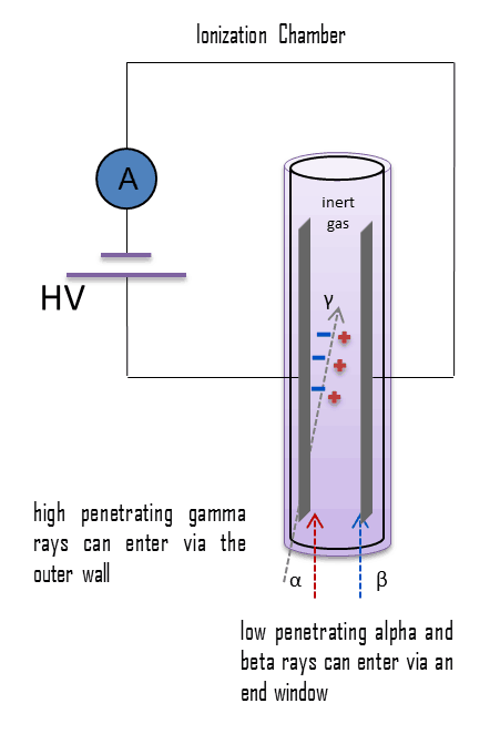
The chamber has a cathode and an anode that are held at some voltage (perhaps 100 – 200 V), and the device is characterized by a capacitance that is determined by the geometry of the electrodes. Flat plates or concentric cylinders may be utilized in the construction of an ionization chamber. The flat plate design is preferred because it has a well-defined active volume and ensures that ions will not collect on the insulators and cause a distortion of the electric field. As ionizing radiation enters the gas between the electrodes, a finite number of ion-pairs are formed. The behavior of the resultant ion-pairs is affected by the potential gradient of the electric field within the gas and the type and pressure of the fill gas. Under the influence of the electric field, the positive ions will move toward the negatively charged electrode (outer cylinder or plate), and the negative ions (electrons) will migrate toward the positive electrode (central wire or plate). The electric field in this region keeps the ions from recombining with the electrons. The collection of these ions will produce a charge on the electrodes and an electrical pulse across the detection circuit. In air, the average energy needed to produce an ion is about 34 eV, therefore a 1 MeV radiation completely absorbed in the detector produces about 3 x 10⁴ pair of ions. However it is a small signal, this signal can be considerably amplified using standard electronics. A current of 1 micro-ampere consists of about 1012 electrons per second.
Ionization chamber construction differs from the proportional counter. The flat plate design is preferred for ionization chambers, or concentric cylinders may be utilized in the construction to allow for the integration of pulses produced by the incident radiation. Proportional counters and Geiger counters usually utilize cylinder and central electrode. The proportional counter would require such exact control of the electric field between the electrodes that it would not be practical.
Detection of Alpha Radiation using Ionization Chamber
For alpha and beta particles to be detected by ionization chambers, they must be provided with a thin window. This “end-window” must be thin enough for the alpha and beta particles to penetrate. However, a window of almost any thickness will prevent an alpha particle from entering the chamber. The window is usually made of mica with a density of about 1.5 – 2.0 mg/cm2. But it does not mean, alpha radiation cannot be detected by an ionization chamber.
For example, in some kind of smoke detectors, you can meet man-made radionuclides such as americium-241, which is a source of alpha particles. The smoke detector has two ionization chambers, one open to the air, and a reference chamber which does not allow the entry of particles. The radioactive source emits alpha particles into both chambers, which ionizes some air molecules. The free-air chamber allows the entry of smoke particles to the sensitive volume and to change attenuation of alpha particles. If any smoke particles enter the free-air chamber, some of the ions will attach to the particles and not be available to carry the current in that chamber. An electronic circuit detects that a current difference has developed between the open and sealed chambers, and sounds the alarm.
Detection of Beta Radiation using Ionization Chamber
For alpha and beta particles to be detected by ionization chambers, they must be provided with a thin window. This “end-window” must be thin enough for the alpha and beta particles to penetrate. However, a window of almost any thickness will prevent an alpha particle from entering the chamber. The window is usually made of mica with a density of about 1.5 – 2.0 mg/cm2.
Ionization chamber may be, for example, used for the measurement of tritium in the air. These devices are known as tritium-in-air monitors. Tritium is a radioactive isotope, but it emits a very weak form of radiation, a low-energy beta particle that is similar to an electron. It is a pure beta emitter (i.e. beta emitter without an accompanying gamma radiation). The electron’s kinetic energy varies, with an average of 5.7 keV, while the remaining energy is carried off by the nearly undetectable electron antineutrino. Such a very low energy of electron causes, that the electron cannot penetrate the skin or even does not travel very far in air. Beta particles from tritium can penetrate only about 6.0 mm of air. It is practically impossible to design a detector whose walls these beta particles can penetrate. Instead, tritium-in-air monitor pumps the tritium-contaminated air right through an ionization chamber, so that all of the energy of the beta particles can be usefully converted to producing ion pairs inside the chamber.
Detection of Gamma Radiation using Ionization Chamber
Gamma rays have very little trouble in penetrating the metal walls of the chamber. Therefore, ionization chambers may be used to detect gamma radiation and X-rays collectively known as photons, and for this the windowless tube is used. Ionization chambers have a good uniform response to radiation over a wide range of energies and are the preferred means of measuring high levels of gamma radiation. Some problems are caused by the fact, that alpha particles are more ionising than beta particles and than gamma rays, so more current is produced in the ionization chamber region by alpha than beta and gamma. Gamma rays deposit significantly lower amount of energy to the detector than other particles.
The efficiency of the chamber can be further increased by the use of a high pressure gas. Typically a pressure of 8-10 atmospheres can be used, and various noble gases are employed. For example, high-pressure xenon (HPXe) ionization chambers are ideal for use in uncontrolled environments, as a detector’s response has been shown to be uniform over large temperature ranges (20–170°C). The higher pressure results in a greater gas density and thereby a greater chance of collision with the fill gas and ion-pair creation by incident gamma radiation. Because of the increased wall thickness required to withstand this high pressure, only gamma radiation can be detected. These detectors are used in survey meters and for environmental monitoring.
Detection of Neutrons using Ionization Chamber
Since the neutrons are electrically neutral particles, they are mainly subject to strong nuclear forces but not to electric forces. Therefore neutrons are not directly ionizing and they have usually to be converted into charged particles before they can be detected. Generally every type of neutron detector must be equipped with converter (to convert neutron radiation to common detectable radiation) and one of the conventional radiation detectors (scintillation detector, gaseous detector, semiconductor detector, etc.).
Ionization chambers are often used as the charged particle detection device. For example, if the inner surface of the ionization chamber is coated with a thin coat of boron, the (n,alpha) reaction can take place. Most of (n,alpha) reactions of thermal neutrons are 10B(n,alpha)7Li reactions accompanied by 0.48 MeV gamma emission.
Moreover, isotope boron-10 has high (n,alpha) reaction cross-section along the entire neutron energy spectrum. The alpha particle causes ionization within the chamber, and ejected electrons cause further secondary ionizations.
Another method for detecting neutrons using an ionization chamber is to use the gas boron trifluoride (BF3) instead of air in the chamber. The incoming neutrons produce alpha particles when they react with the boron atoms in the detector gas. Either method may be used to detect neutrons in nuclear reactor. It must be noted, BF3 counters are usually operated in the proportional region.
Robert Reed Burn, Introduction to Nuclear Reactor Operation, 1988.
U.S. Department of Energy, Nuclear Physics and Reactor Theory. DOE Fundamentals Handbook, Volume 1 and 2. January 1993.
Paul Reuss, Neutron Physics. EDP Sciences, 2008. ISBN: 978-2759800414.
See also:
Gaseous Detectors
We hope, this article, Ionization Chamber – Ion Chamber, helps you. If so, give us a like in the sidebar. Main purpose of this website is to help the public to learn some interesting and important information about radiation and dosimeters.
Measurement of Radiation. Detailed knowledge about detection of radiation is very important in many branches of engineering, including radiation protection. Radiation Dosimetry
Source of gamma radiation. The danger of ionizing radiation lies in the fact that the radiation is invisible and not directly detectable by human senses.
The danger of ionizing radiation lies in the fact that the radiation is invisible and not directly detectable by human senses. People can neither see nor feel radiation, yet it deposits energy to the molecules of material. The energy is transferred in small quantities for each interaction between the radiation and a molecule and there are usually many types of interactions. Therefore, the only way you can detect and measure radiation is to use instruments (detectors of ionizing radiation).
Detailed knowledge about detection of radiation is very important in many branches of engineering, including radiation protection. Most modern nuclear or particle experiments use a variety of sophisticated detectors for measuring and detection of sub-atomic particles. In order to be detected, a particle must leave some trace of its presence in a detector. Particles mostly deposit energy along its path. Knowledge of this interaction, how different particles deposit energy in the matter and how much energy particles deposit, is fundamental for our understanding of many problems. This chapter will give you a basic understanding of how these detectors work and some of their limitations.
What is Ionization
Ionization is the process in which an atom or a molecule gains or loses electrons to form charged ion. Ionization can result from the loss of an electron after collisions with energetic subatomic particles, collisions with other atoms, molecules and ions, or through the interaction with electromagnetic radiation. In general, ionizing radiation is any radiation (particles or electromagnetic waves) that carries enough energy to knock electrons from atoms or molecules, thereby ionizing them. For ionizing radiation, the kinetic energy of particles (photons, electrons, etc.) is sufficient and the particle can ionize (to form ion by losing electrons) target atoms to form ions.
The boundary between ionizing and non-ionizing radiation is not sharply defined, since different molecules and atoms ionize at different energies. Gamma rays, X-rays, and the higher ultraviolet part of the spectrum are ionizing, whereas the lower ultraviolet, visible light (including laser light), infrared, microwaves, and radio waves are considered non-ionizing radiation.
Basic Principles of Detectors
Detectors of ionizing radiation consist of two parts that are usually connected. The first part consists of a sensitive material, consisting of a compound that experiences changes when exposed to radiation. The other component is a device that converts these changes into measurable signals.
There are three main types of detectors, which record different types of signals.
Counter. The activity or intensity of radiation is measured in counts per second (cps). The best known counter is the Geiger-Müller counter. In radiation counters the generated signal from the incident radiation is created by the counting of the number of interactions occurring at the sensitive volume of the detector.
Radiation Spectrometer. Spectrometers are devices designed to measure the spectral power distribution of a source. The incident radiation generates a signal that allows to determine the energy of the incident particle.
Dosimeter. A radiation dosimeter is a device that measures exposure to ionizing radiation. Dosimeters usually record a dose, which is the absorbed radiation energy measured in grays (Gy) of the equivalent dose measured in sieverts (Sv) A personal dosimeter is dosimeter, that is worn at the surface of the body by the person being monitored, and it records of the radiation dose received.
All these types of equipment require that the radiations result in observable changes in a compound (whether gas, liquid or solid). In their basic principles of operation, most detectors of ionizing radiation follow similar characteristics. Detectors of ionizing radiation consist of two parts that are usually connected. The first part consists of a sensitive material, consisting of a compound that experiences changes when exposed to radiation. The other component is a device that converts these changes into measurable signals. All detectors require that radiation must deposit some of its energy in sensitive material that forms part of the instrument. The radiation enters the detector, interacts with atoms of the detector material and deposits some energy to sensitive material. Each event may generate a signal, which can be a pulse, hole, light signal, ion pairs in a gas, and many others. The main task is to generate sufficient signal, amplify it and to record it.
Let assume gaseous ionization detectors. Basic gaseous ionization detector consists of a chamber that is filled with a suitable medium (air or a special fill gas) that can be easily ionized. As a general rule, the center wire is the positive electrode (anode) and the outer cylinder is the negative electrode (cathode), so that (negative) electrons are attracted to the center wire and positive ions are attracted to the outer cylinder. The anode is at a positive voltage with respect to the detector wall. As ionizing radiation enters the gas between the electrodes, a finite number of ion-pairs are formed. Under the influence of the electric field, the positive ions will move toward the negatively charged electrode (outer cylinder), and the negative ions (electrons) will migrate toward the positive electrode (central wire). The collection of these ions will produce a charge on the electrodes and an electrical pulse across the detection circuit. However it is a small signal, this signal can be amplified, and then recorded using standard electronics.
Robert Reed Burn, Introduction to Nuclear Reactor Operation, 1988.
U.S. Department of Energy, Nuclear Physics and Reactor Theory. DOE Fundamentals Handbook, Volume 1 and 2. January 1993.
Paul Reuss, Neutron Physics. EDP Sciences, 2008. ISBN: 978-2759800414.
See also:
Radiation Detection
We hope, this article, Measurement of Radiation, helps you. If so, give us a like in the sidebar. Main purpose of this website is to help the public to learn some interesting and important information about radiation and dosimeters.
Ionization is the process in which an atom or a molecule gains or loses electrons to form charged ion. Ionization can result from the loss of an electron after collisions with energetic subatomic particles. Radiation Dosimetry
What is Ionization
Ionization is the process in which an atom or a molecule gains or loses electrons to form charged ion. Ionization can result from the loss of an electron after collisions with energetic subatomic particles, collisions with other atoms, molecules and ions, or through the interaction with electromagnetic radiation. In general, ionizing radiation is any radiation (particles or electromagnetic waves) that carries enough energy to knock electrons from atoms or molecules, thereby ionizing them. For ionizing radiation, the kinetic energy of particles (photons, electrons, etc.) is sufficient and the particle can ionize (to form ion by losing electrons) target atoms to form ions.
The boundary between ionizing and non-ionizing radiation is not sharply defined, since different molecules and atoms ionize at different energies. Gamma rays, X-rays, and the higher ultraviolet part of the spectrum are ionizing, whereas the lower ultraviolet, visible light (including laser light), infrared, microwaves, and radio waves are considered non-ionizing radiation.
Ionization Energy
Ionization energy, also called ionization potential, is the energy necessary to remove an electron from the neutral atom.
X + energy → X+ + e−
where X is any atom or molecule capable of being ionized, X+ is that atom or molecule with an electron removed (positive ion), and e− is the removed electron.
A nitrogen atom, for example, requires the following ionization energy to remove the outermost electron.
N + IE → N+ + e− IE = 14.5 eV
The ionization energy associated with removal of the first electron is most commonly used. The nth ionization energy refers to the amount of energy required to remove an electron from the species with a charge of (n-1).
1st ionization energy
X → X+ + e−
2nd ionization energy
X+ → X2+ + e−
3rd ionization energy
X2+ → X3+ + e−
Ionization Energy for different Elements
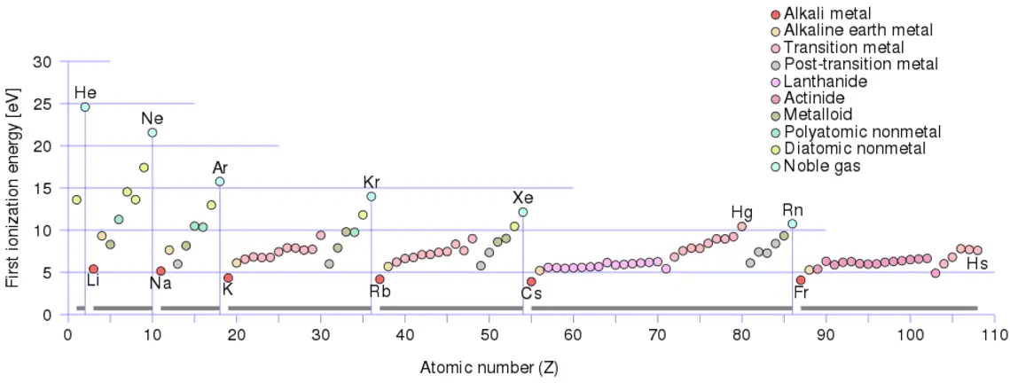
There is an ionization energy for each successive electron removed. The electrons that circle the nucleus move in fairly well-defined orbits. Some of these electrons are more tightly bound in the atom than others. For example, only 7.38 eV is required to remove the outermost electron from a lead atom, while 88,000 eV is required to remove the innermost electron. Helps to understand reactivity of elements (especially metals, which lose electrons).
In general, the ionization energy increases moving up a group and moving left to right across a period. Moreover:
Ionization energy is lowest for the alkali metals which have a single electron outside a closed shell.
Ionization energy increases across a row on the periodic maximum for the noble gases which have closed shells
For example, sodium requires only 496 kJ/mol or 5.14 eV/atom to ionize it. On the other hand neon, the noble gas, immediately preceding it in the periodic table, requires 2081 kJ/mol or 21.56 eV/atom.
Robert Reed Burn, Introduction to Nuclear Reactor Operation, 1988.
U.S. Department of Energy, Nuclear Physics and Reactor Theory. DOE Fundamentals Handbook, Volume 1 and 2. January 1993.
Paul Reuss, Neutron Physics. EDP Sciences, 2008. ISBN: 978-2759800414.
See also:
Radiation Detection
We hope, this article, Ionization, helps you. If so, give us a like in the sidebar. Main purpose of this website is to help the public to learn some interesting and important information about radiation and dosimeters.
In their basic principles of operation, most detectors of ionizing radiation follow similar characteristics. Detectors of ionizing radiation consist of two parts that are usually connected. Radiation Dosimetry
Basic Principles of Detectors
Detectors of ionizing radiation consist of two parts that are usually connected. The first part consists of a sensitive material, consisting of a compound that experiences changes when exposed to radiation. The other component is a device that converts these changes into measurable signals.
There are three main types of detectors, which record different types of signals.
Counter. The activity or intensity of radiation is measured in counts per second (cps). The best known counter is the Geiger-Müller counter. In radiation counters the generated signal from the incident radiation is created by the counting of the number of interactions occurring at the sensitive volume of the detector.
Radiation Spectrometer. Spectrometers are devices designed to measure the spectral power distribution of a source. The incident radiation generates a signal that allows to determine the energy of the incident particle.
Dosimeter. A radiation dosimeter is a device that measures exposure to ionizing radiation. Dosimeters usually record a dose, which is the absorbed radiation energy measured in grays (Gy) of the equivalent dose measured in sieverts (Sv) A personal dosimeter is dosimeter, that is worn at the surface of the body by the person being monitored, and it records of the radiation dose received.
All these types of equipment require that the radiations result in observable changes in a compound (whether gas, liquid or solid). In their basic principles of operation, most detectors of ionizing radiation follow similar characteristics. Detectors of ionizing radiation consist of two parts that are usually connected. The first part consists of a sensitive material, consisting of a compound that experiences changes when exposed to radiation. The other component is a device that converts these changes into measurable signals. All detectors require that radiation must deposit some of its energy in sensitive material that forms part of the instrument. The radiation enters the detector, interacts with atoms of the detector material and deposits some energy to sensitive material. Each event may generate a signal, which can be a pulse, hole, light signal, ion pairs in a gas, and many others. The main task is to generate sufficient signal, amplify it and to record it.
In this section we describe a number of different types of radiation measuring instruments. We focus on the first part, which consists of a sensitive material. Detectors may be categorized according to sensitive materials and methods that can be utilized to make a measurement:
Gaseous Ionization Detectors
Scintillation Detectors
Semiconductor Detectors
Film Badges
TLD
Let assume gaseous ionization detectors. Basic gaseous ionization detector consists of a chamber that is filled with a suitable medium (air or a special fill gas) that can be easily ionized. As a general rule, the center wire is the positive electrode (anode) and the outer cylinder is the negative electrode (cathode), so that (negative) electrons are attracted to the center wire and positive ions are attracted to the outer cylinder. The anode is at a positive voltage with respect to the detector wall. As ionizing radiation enters the gas between the electrodes, a finite number of ion-pairs are formed. Under the influence of the electric field, the positive ions will move toward the negatively charged electrode (outer cylinder), and the negative ions (electrons) will migrate toward the positive electrode (central wire). The collection of these ions will produce a charge on the electrodes and an electrical pulse across the detection circuit. However it is a small signal, this signal can be amplified, and then recorded using standard electronics.
Robert Reed Burn, Introduction to Nuclear Reactor Operation, 1988.
U.S. Department of Energy, Nuclear Physics and Reactor Theory. DOE Fundamentals Handbook, Volume 1 and 2. January 1993.
Paul Reuss, Neutron Physics. EDP Sciences, 2008. ISBN: 978-2759800414.
See also:
Radiation Detection
We hope, this article, Principle of Detectors – Method of Detection, helps you. If so, give us a like in the sidebar. Main purpose of this website is to help the public to learn some interesting and important information about radiation and dosimeters.
There are many types of radiation detectors, which record different types of signals. Classification of radiation detectors may be done in many ways. Radiation Dosimetry
Purpose of Radiation Detectors
Detectors of ionizing radiation consist of two parts that are usually connected. The first part consists of a sensitive material, consisting of a compound that experiences changes when exposed to radiation. The other component is a device that converts these changes into measurable signals.
There are three main types of detectors, which record different types of signals.
Counter. The activity or intensity of radiation is measured in counts per second (cps). The best known counter is the Geiger-Müller counter. In radiation counters the generated signal from the incident radiation is created by the counting of the number of interactions occurring at the sensitive volume of the detector.
Radiation Spectrometer. Spectrometers are devices designed to measure the spectral power distribution of a source. The incident radiation generates a signal that allows to determine the energy of the incident particle.
Dosimeter. A radiation dosimeter is a device that measures exposure to ionizing radiation. Dosimeters usually record a dose, which is the absorbed radiation energy measured in grays (Gy) of the equivalent dose measured in sieverts (Sv) A personal dosimeter is dosimeter, that is worn at the surface of the body by the person being monitored, and it records of the radiation dose received.
All these types of equipment require that the radiations result in observable changes in a compound (whether gas, liquid or solid). In their basic principles of operation, most detectors of ionizing radiation follow similar characteristics. Detectors of ionizing radiation consist of two parts that are usually connected. The first part consists of a sensitive material, consisting of a compound that experiences changes when exposed to radiation. The other component is a device that converts these changes into measurable signals. All detectors require that radiation must deposit some of its energy in sensitive material that forms part of the instrument. The radiation enters the detector, interacts with atoms of the detector material and deposits some energy to sensitive material. Each event may generate a signal, which can be a pulse, hole, light signal, ion pairs in a gas, and many others. The main task is to generate sufficient signal, amplify it and to record it.
Classification by Sensitive Material
We focus on the first part, which consists of a sensitive material. Detectors may be categorized according to sensitive materials and methods that can be utilized to make a measurement:
Gaseous Ionization Detectors
Scintillation Detectors
Semiconductor Detectors
Film Badges
TLD
Let assume gaseous ionization detectors. Basic gaseous ionization detector consists of a chamber that is filled with a suitable medium (air or a special fill gas) that can be easily ionized. As a general rule, the center wire is the positive electrode (anode) and the outer cylinder is the negative electrode (cathode), so that (negative) electrons are attracted to the center wire and positive ions are attracted to the outer cylinder. The anode is at a positive voltage with respect to the detector wall. As ionizing radiation enters the gas between the electrodes, a finite number of ion-pairs are formed. Under the influence of the electric field, the positive ions will move toward the negatively charged electrode (outer cylinder), and the negative ions (electrons) will migrate toward the positive electrode (central wire). The collection of these ions will produce a charge on the electrodes and an electrical pulse across the detection circuit. However it is a small signal, this signal can be amplified, and then recorded using standard electronics.
Classification of Radiation Detectors – Radiation Type
Principles and methods for detection of ionizing radiation are dependent on many factors. Type of radiation measured and detected is a key factor, and different types of detectors in different physical states (solid, liquid or gas) are used to measure selective types of ionizing radiation. It is important to distinguish between alpha or beta particles, X-rays or γ-rays and neutrons. Since each radiation interacts differently with matter, we cannot use, for example, neutron detector to detect beta radiation. Sometimes, detectors can detect various types of radiation, for example, neutron detectors are also able to detect gamma radiation. Usually, this feature is not desirable, and detectors must compensate the undesirable component of radiation.
In order to describe the classification of radiation detectors by the type of radiation, we have to understand the interactions of radiation with matter. Each type particle interacts in a different way, therefore we must describe interaction of particles (radiation as a flow of these particles) separately. For example charged particles with high energies can directly ionize atoms. On the other hand electrically neutral particles interacts only indirectly, but can also transfer some or all of their energies to the matter. This is the key feature of the categorization of radiation detectors. Simply, we cannot use a gamma rays detector to detect alpha radiation. Detectors can be categorized into two general types as follows:
Robert Reed Burn, Introduction to Nuclear Reactor Operation, 1988.
U.S. Department of Energy, Nuclear Physics and Reactor Theory. DOE Fundamentals Handbook, Volume 1 and 2. January 1993.
Paul Reuss, Neutron Physics. EDP Sciences, 2008. ISBN: 978-2759800414.
See also:
Radiation Detection
We hope, this article, Classification of Radiation Detectors – Types of Radiation Detectors, helps you. If so, give us a like in the sidebar. Main purpose of this website is to help the public to learn some interesting and important information about radiation and dosimeters.
 Fast Fission. The fast fission process is in the multiplication factor characterized by the fast fission factor, ε, which increases the fast neutron population in one neutron generation. The fast fission factor is defined as the ratio of the fast neutrons produced by fissions at all energies to the number of fast neutrons produced in thermal fission.