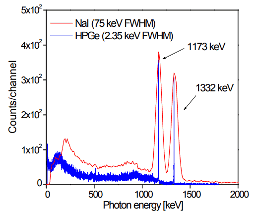
Analysis of gamma spectra is very interesting, since it has a structure and workers must distinguish between true pulses to be analyzed and accompanying pulses from different sources of radiation. We will show the structure of the gamma spectrum on the example of cobalt-60 measured by NaI(Tl) scintillation detector and by the HPGe detector. The HPGe detector allows the separation of many closely spaced gamma lines, which is very beneficial for measuring multi-gamma emitting radioactive sources.

Cobalt-60 is an artificial radioactive isotope of cobalt with a half-life of 5.2747 years. It is synthetically produced by neutron activation of cobalt-59 in nuclear reactors. Cobalt-60 is a common calibration source found in many laboratories. The gamma spectrum has two significant peaks, one at 1173.2 keV and another at 1332.5 keV. Good scintillation detectors should have adequate resolution to separate the two peaks. For HPGe detectors, these peaks are perfectly separated.
As can be seen from the figure, there are two gamma ray photopeaks. Both detectors also show response at the lower energies, caused by Compton scattering, two smaller escape peaks at energies 0.511 and 1.022 MeV below the photopeak for the creation of electron-positron pairs when one or both annihilation photons escape, and a backscatter peak. Higher energies can be measured when two or more photons strike the detector almost simultaneously, appearing as sum peaks with energies up to the value of two or more photopeaks added.
X-ray Peaks
When the gamma rays undergoes photoelectric effect in surrounding materials (for example lead shield) the outgoing X-ray can be captured again by the detector. This gives an characteristic X-ray peak with an energy depending on the material it came from. In case of lead the characteristic X-ray energies is in the 72-84 keV range. Photoelectric absorption by K-shell electron in lead of shielding, resulting in a K-shell vacancy. The K -> L transition for lead = 72 keV. If this characteristic x-ray is absorbed in the crystal then a secondary peak at 72 keV is observed.
We hope, this article, X-Ray Peak – Spectrum, helps you. If so, give us a like in the sidebar. Main purpose of this website is to help the public to learn some interesting and important information about radiation and dosimeters.