Detection of Directly Ionizing Radiation. Charged particles (atomic nuclei, electrons, positrons, protons, muons, etc.) can ionize atoms directly by fundamental interaction through the Coulomb force. Radiation Dosimetry
Charged particles (atomic nuclei, electrons, positrons, protons, muons, etc.) can ionize atoms directly by fundamental interaction through the Coulomb force if it carries sufficient kinetic energy. These particles must be moving at relativistic speeds to reach the required kinetic energy. Even photons (gamma rays and X-rays) can ionize atoms directly (despite they are electrically neutral) through the Photoelectric effect and the Compton effect, but secondary (indirect) ionization is much more significant.
Detection of Alpha Particles
Detection of alpha radiation is very specific, because alpha particles travel only a few centimeters in air but deposit all their energies along their short paths, thus the amount of energy transferred is very high.
In order to describe principles of detection of alpha radiation, we have to understand the interaction of radiation with matter. Each type particle interacts in a different way, therefore we must describe interaction of alpha particles (radiation as a flow of these particles) separately.
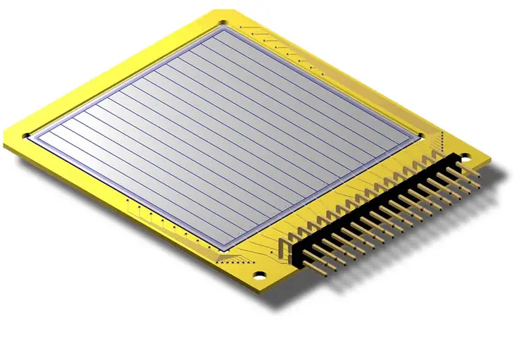
Detection of Alpha using Semiconductors – Silicon Strip Detectors
Silicon-based detectors are very good for tracking charged particles. A silicon strip detector is an arrangement of strip like shaped implants acting as charge collecting electrodes.
Silicon strip detectors 5 x 5 cm2 in area are quite common and are used in series (just like planes of MWPCs) to determine charged-particle trajectories to position-accuracies of the order of several μm in the transverse direction. Placed on a low doped fully depleted silicon wafer these implants form a one-dimensional array of diodes. By connecting each of the metalized strips to a charge sensitive amplifier a position sensitive detector is built. Two dimensional position measurements can be achieved by applying an additional strip like doping on the wafer backside by use of a double sided technology. Such devices can be used to measure small impact parameters and thereby determine whether some charged particle originated from a primary collision or was the decay product of a primary particle that traveled a small distance from the original interaction, and then decayed.
Detection of Beta Particles
Detection of beta radiation is very specific, because beta particles are more penetrating than alpha particles. On the other hand a thin aluminum plate can stop them.
In order to describe principles of detection of beta radiation, we have to understand the interaction of radiation with matter. Each type particle interacts in a different way, therefore we must describe interaction of beta particles (radiation as a flow of these particles) separately.
Detection of Beta Radiation using Scintillation Counter
Scintillation counters are used to measure radiation in a variety of applications including hand held radiation survey meters, personnel and environmental monitoring for radioactive contamination, medical imaging, radiometric assay, nuclear security and nuclear plant safety. They are widely used because they can be made inexpensively yet with good efficiency, and can measure both the intensity and the energy of incident radiation.
Beta Particles. For detection of beta particles, organic scintillators can be used. Pure organic crystals include crystals of anthracene, stilbene and naphthalene. The decay time of this type of phosphor is approximately 10 nanoseconds. This type of crystal is frequently used in the detection of beta particles. Organic scintillators, having a lower Z than inorganic crystals, are best suited for the detection of low-energy (< 10 MeV) beta particles.
Robert Reed Burn, Introduction to Nuclear Reactor Operation, 1988.
U.S. Department of Energy, Nuclear Physics and Reactor Theory. DOE Fundamentals Handbook, Volume 1 and 2. January 1993.
Paul Reuss, Neutron Physics. EDP Sciences, 2008. ISBN: 978-2759800414.
See also:
Detectors
We hope, this article, Detection of Directly Ionizing Radiation, helps you. If so, give us a like in the sidebar. Main purpose of this website is to help the public to learn some interesting and important information about radiation and dosimeters.
Detection of indirectly ionizing radiation is very specific, because, indirect ionizing radiation consist of electrically neutral particles and therefore does not ionize matter directly. Radiation Dosimetry
Indirect ionizing radiation consist of electrically neutral particles and therefore does not ionize matter directly. The bulk of the ionization effects are due to secondary ionizations.
Detection of Gamma Radiation
Detection of gamma radiation is very specific, because gamma rays interact differently with matter. Gamma rays can travel thousands of feet in air and can easily pass through various materials. Moreover, gamma rays can ionize atoms indirectly and directly (despite they are electrically neutral) through the photoelectric effect and the Compton effect. But secondary (indirect) ionization is much more significant.
In order to describe principles of detection of gamma radiation, we have to understand the interaction of radiation with matter. Each type particle interacts in a different way, therefore we must describe interactions of gamma rays (radiation as a flow of these rays) separately.
Detection of Gamma Radiation using Ionization Chamber
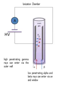
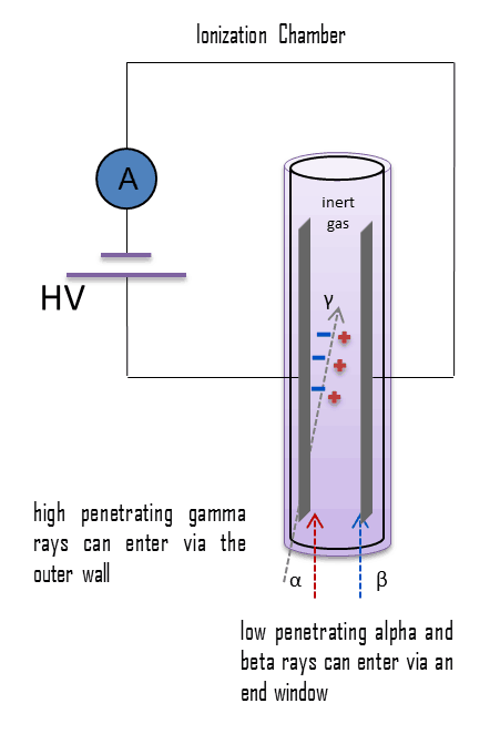
Gamma rays have very little trouble in penetrating the metal walls of the chamber. Therefore, ionization chambers may be used to detect gamma radiation and X-rays collectively known as photons, and for this the windowless tube is used. Ionization chambers have a good uniform response to radiation over a wide range of energies and are the preferred means of measuring high levels of gamma radiation. Some problems are caused by the fact, that alpha particles are more ionising than beta particles and than gamma rays, so more current is produced in the ionization chamber region by alpha than beta and gamma. Gamma rays deposit significantly lower amount of energy to the detector than other particles.
The efficiency of the chamber can be further increased by the use of a high pressure gas. Typically a pressure of 8-10 atmospheres can be used, and various noble gases are employed. For example, high-pressure xenon (HPXe) ionization chambers are ideal for use in uncontrolled environments, as a detector’s response has been shown to be uniform over large temperature ranges (20–170°C). The higher pressure results in a greater gas density and thereby a greater chance of collision with the fill gas and ion-pair creation by incident gamma radiation. Because of the increased wall thickness required to withstand this high pressure, only gamma radiation can be detected. These detectors are used in survey meters and for environmental monitoring.
Detection of Neutrons
Generally every type of neutron detector must be equipped with converter and one of the conventional radiation detectors. Source: large.stanford.edu
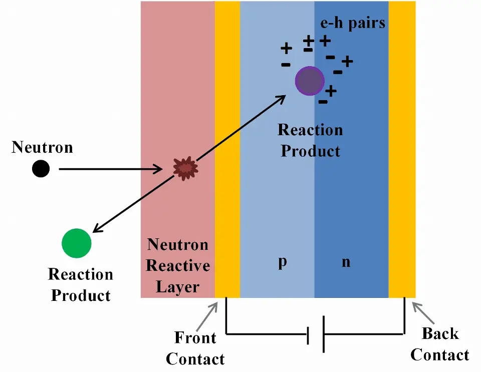
Detection of neutrons is very specific, since the neutrons are electrically neutral particles, thus they are mainly subject to strong nuclear forces but not to electric forces. Therefore neutrons are not directly ionizing and they have usually to be converted into charged particles before they can be detected. Generally every type of neutron detector must be equipped with converter (to convert neutron radiation to common detectable radiation) and one of the conventional radiation detectors (scintillation detector, gaseous detector, semiconductor detector, etc.).
Detection of Neutrons using Ionization Chamber
Ionization chambers are often used as the charged particle detection device. For example, if the inner surface of the ionization chamber is coated with a thin coat of boron, the (n,alpha) reaction can take place. Most of (n,alpha) reactions of thermal neutrons are 10B(n,alpha)7Li reactions accompanied by 0.48 MeV
Moreover, isotope boron-10 has high (n,alpha) reaction cross-section along the entire neutron energy spectrum. The alpha particle causes ionization within the chamber, and ejected electrons cause further secondary ionizations.
Another method for detecting neutrons using an ionization chamber is to use the gas boron trifluoride (BF3) instead of air in the chamber. The incoming neutrons produce alpha particles when they react with the boron atoms in the detector gas. Either method may be used to detect neutrons in nuclear reactor. It must be noted, BF3 counters are usually operated in the proportional region.
Robert Reed Burn, Introduction to Nuclear Reactor Operation, 1988.
U.S. Department of Energy, Nuclear Physics and Reactor Theory. DOE Fundamentals Handbook, Volume 1 and 2. January 1993.
Paul Reuss, Neutron Physics. EDP Sciences, 2008. ISBN: 978-2759800414.
See also:
Detectors
We hope, this article, Detection of Indirectly Ionizing Radiation, helps you. If so, give us a like in the sidebar. Main purpose of this website is to help the public to learn some interesting and important information about radiation and dosimeters.
Detection of beta radiation is very specific, because beta particles are more penetrating than alpha particles. On the other hand a thin aluminum plate can stop them. Detection of Beta Radiation – Beta Particle Detector
Laboratory use of a Geiger counter with end-window probe to measure beta radiation Source: wikipedia.org License: Public Domain
Detection of beta radiation is very specific, because beta particles are more penetrating than alpha particles. On the other hand a thin aluminum plate can stop them.
In order to describe principles of detection of beta radiation, we have to understand the interaction of radiation with matter. Each type particle interacts in a different way, therefore we must describe interaction of beta particles (radiation as a flow of these particles) separately.
Interaction of Beta Particles with Matter
Beta radiation consist of free electrons or positrons at relativistic speeds. Beta particles (electrons) are much smaller than alpha particles. They carry a single negative charge. They are more penetrating than alpha particles, but a thin aluminium plate can stop them. They can travel several meters in air but deposit less energy at any one point along their paths than alpha particles. Similarly as for heavy charged particles, beta particles transfer energy by:
Excitation. The charged particle can transfer energy to the atom, raising electrons to a higher energy levels.
Ionization. Ionization can occur, when the charged particle have enough energy to remove an electron. This results in a creation of ion pairs in surrounding matter.
In addition to these interactions, beta particles also lose energy by radiative process known as the bremsstrahlung. From classical theory, when a charged particle is accelerated or decelerated, it must radiate energy and the deceleration radiation is known as the bremsstrahlung (“braking radiation”).
Summary of types of interactions:
Inelastic collisions with atomic electrons (Excitation and Ionization)
Nature of an interaction of a beta radiation with matter is different from the alpha radiation, despite the fact that beta particles are also charged particles. In comparison with alpha particles, beta particles have much lower mass and they reach mostly relativistic energies. Their mass is equal to the mass of the orbital electrons with which they are interacting and unlike the alpha particle a much larger fraction of its kinetic energy can be lost in a single interaction. Since the beta particles mostly reach relativistic energies, the nonrelativistic Bethe formula cannot be used. For high energy electrons an similar expression has also been derived by Bethe to describe the specific energy loss due to excitation and ionization (the “collisional losses”).
Moreover, beta particles can interact via electron-nuclear interaction (elastic scattering off nuclei), which can significantly change the direction of beta particle. Therefore their path is not so straightforward. The beta particles follow a very zig-zag path through absorbing material, this resulting path of particle is longer than the linear penetration (range) into the material.
Detectors of Beta Radiation
Detectors may be also categorized according to sensitive materials and methods that can be utilized to make a measurement:
Detection of Beta Radiation using Ionization Chamber
For alpha and beta particles to be detected by ionization chambers, they must be provided with a thin window. This “end-window” must be thin enough for the alpha and beta particles to penetrate. However, a window of almost any thickness will prevent an alpha particle from entering the chamber. The window is usually made of mica with a density of about 1.5 – 2.0 mg/cm2.
Ionization chamber may be, for example, used for the measurement of tritium in the air. These devices are known as tritium-in-air monitors. Tritium is a radioactive isotope, but it emits a very weak form of radiation, a low-energy beta particle that is similar to an electron. It is a pure beta emitter (i.e. beta emitter without an accompanying gamma radiation). The electron’s kinetic energy varies, with an average of 5.7 keV, while the remaining energy is carried off by the nearly undetectable electron antineutrino. Such a very low energy of electron causes, that the electron cannot penetrate the skin or even does not travel very far in air. Beta particles from tritium can penetrate only about 6.0 mm of air. It is practically impossible to design a detector whose walls these beta particles can penetrate. Instead, tritium-in-air monitor pumps the tritium-contaminated air right through an ionization chamber, so that all of the energy of the beta particles can be usefully converted to producing ion pairs inside the chamber.
Detection of Beta Radiation using Scintillation Counter
Scintillation counters are used to measure radiation in a variety of applications including hand held radiation survey meters, personnel and environmental monitoring for radioactive contamination, medical imaging, radiometric assay, nuclear security and nuclear plant safety. They are widely used because they can be made inexpensively yet with good efficiency, and can measure both the intensity and the energy of incident radiation.
Beta Particles. For detection of beta particles, organic scintillators can be used. Pure organic crystals include crystals of anthracene, stilbene and naphthalene. The decay time of this type of phosphor is approximately 10 nanoseconds. This type of crystal is frequently used in the detection of beta particles. Organic scintillators, having a lower Z than inorganic crystals, are best suited for the detection of low-energy (< 10 MeV) beta particles.
Detection of Beta Radiation using Semiconductors – Silicon Strip Detectors
Silicon-based detectors are very good for tracking charged particles. A silicon strip detector is an arrangement of strip like shaped implants acting as charge collecting electrodes.
Silicon strip detectors 5 x 5 cm2 in area are quite common and are used in series (just like planes of MWPCs) to determine charged-particle trajectories to position-accuracies of the order of several μm in the transverse direction. Placed on a low doped fully depleted silicon wafer these implants form a one-dimensional array of diodes. By connecting each of the metalized strips to a charge sensitive amplifier a position sensitive detector is built. Two dimensional position measurements can be achieved by applying an additional strip like doping on the wafer backside by use of a double sided technology. Such devices can be used to measure small impact parameters and thereby determine whether some charged particle originated from a primary collision or was the decay product of a primary particle that traveled a small distance from the original interaction, and then decayed.
Robert Reed Burn, Introduction to Nuclear Reactor Operation, 1988.
U.S. Department of Energy, Nuclear Physics and Reactor Theory. DOE Fundamentals Handbook, Volume 1 and 2. January 1993.
Paul Reuss, Neutron Physics. EDP Sciences, 2008. ISBN: 978-2759800414.
See also:
Detectors
We hope, this article, Detection of Beta Radiation – Beta Particle Detector, helps you. If so, give us a like in the sidebar. Main purpose of this website is to help the public to learn some interesting and important information about radiation and dosimeters.
Detection of gamma radiation is very specific, because gamma rays interact differently with matter. Detection of Gamma Radiation – Detector of Gamma Rays
Detection of gamma radiation is very specific, because gamma rays interact differently with matter. Gamma rays can travel thousands of feet in air and can easily pass through various materials. Moreover, gamma rays can ionize atoms indirectly and directly (despite they are electrically neutral) through the photoelectric effect and the Compton effect. But secondary (indirect) ionization is much more significant.
In order to describe principles of detection of gamma radiation, we have to understand the interaction of radiation with matter. Each type particle interacts in a different way, therefore we must describe interactions of gamma rays (radiation as a flow of these rays) separately.
Interaction of Gamma Radiation with Matter
Gamma rays consist of high energy photons. These photons are particles/waves (Wave-Particle Duality) without rest mass or electrical charge. They can travel 10 meters or more in air. This is a long distance compared to alpha or beta particles. However, gamma rays deposit less energy along their paths. Lead, water, and concrete stop gamma radiation. Photons (gamma rays and X-rays) can ionize atoms directly through the Photoelectric effect and the Compton effect, where the relatively energetic electron is produced. The secondary electron will go on to produce multiple ionization events, therefore the secondary (indirect) ionization is much more significant.
Although a large number of possible interactions are known, there are three key interaction mechanisms with matter.
The photon is completely absorbed in photoelectric effect and pair production, while only partial energy is deposited in any given Compton scattering. The probability of photoelectric absorption (dominates at lower gamma rays energies) per unit mass is approximately proportional to:
τ(photoelectric) = constant x ZN/E3.5
where Z is the atomic number, the exponent n varies between 4 and 5. E is the energy of the incident photon. The probability of Compton scattering per one interaction with an atom increases linearly with atomic number Z, because it depends on the number of electrons, which are available for scattering in the target atom. The probability of pair production (dominates at higher gamma rays energies), characterized by cross section, is a very complicated function based on quantum mechanics. In general the cross section increases approximately with the square of atomic number (σp ~ Z2) and increases with photon energy, but this dependence is much more complex.
As a result, effective sensitive material for gamma radiation detection is in most cases based on use of materials with two following material properties:
high-density of material.
high atomic number of material (high Z materials)
Detectors of Gamma Radiation
Detectors may be also categorized according to sensitive materials and methods that can be utilized to make a measurement:
Detection of Gamma Radiation using Ionization Chamber
Gamma rays have very little trouble in penetrating the metal walls of the chamber. Therefore, ionization chambers may be used to detect gamma radiation and X-rays collectively known as photons, and for this the windowless tube is used. Ionization chambers have a good uniform response to radiation over a wide range of energies and are the preferred means of measuring high levels of gamma radiation. Some problems are caused by the fact, that alpha particles are more ionising than beta particles and than gamma rays, so more current is produced in the ionization chamber region by alpha than beta and gamma. Gamma rays deposit significantly lower amount of energy to the detector than other particles.
The efficiency of the chamber can be further increased by the use of a high pressure gas. Typically a pressure of 8-10 atmospheres can be used, and various noble gases are employed. For example, high-pressure xenon (HPXe) ionization chambers are ideal for use in uncontrolled environments, as a detector’s response has been shown to be uniform over large temperature ranges (20–170°C). The higher pressure results in a greater gas density and thereby a greater chance of collision with the fill gas and ion-pair creation by incident gamma radiation. Because of the increased wall thickness required to withstand this high pressure, only gamma radiation can be detected. These detectors are used in survey meters and for environmental monitoring.
Detection of Gamma Radiation using Geiger Counter
Detector of Ionizing Radiation – Geiger Tube
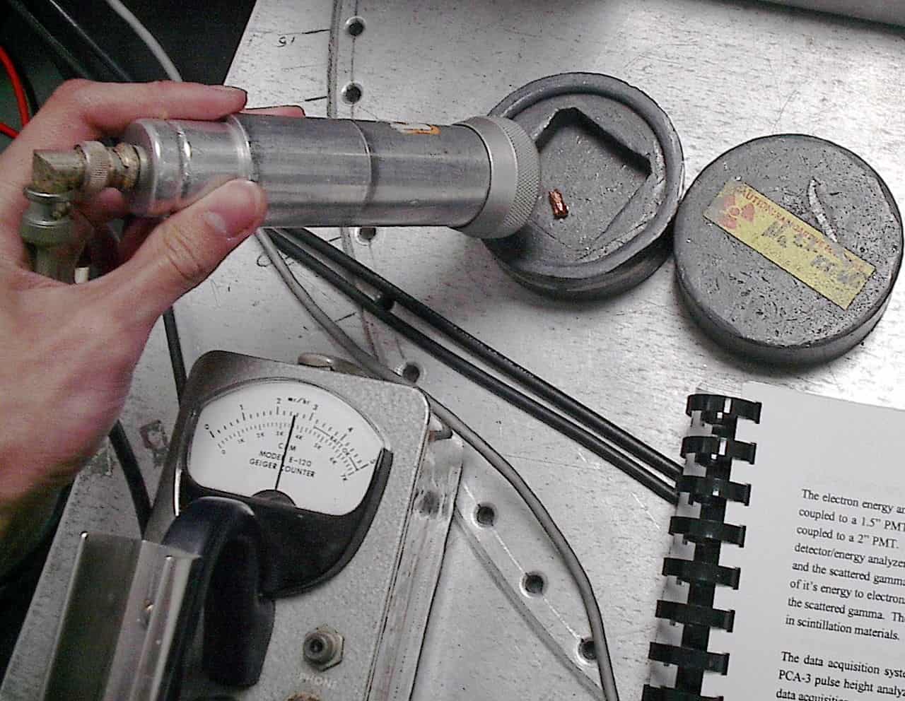
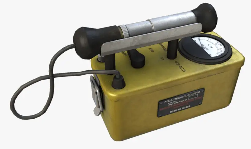
Geiger counter can detect ionizing radiation such as alpha and beta particles, neutrons, and gamma rays using the ionization effect produced in a Geiger–Müller tube, which gives its name to the instrument. The voltage of detector is adjusted so that the conditions correspond to the Geiger-Mueller region.
The high amplification factor of the Geiger counter is the major advantage over the ionization chamber. Geiger counter is therefore a much more sensitive device than other chambers. It is often used in the detection of low-level gamma rays and beta particles for this reason.
Detection of Gamma Radiation using Scintillation Counter
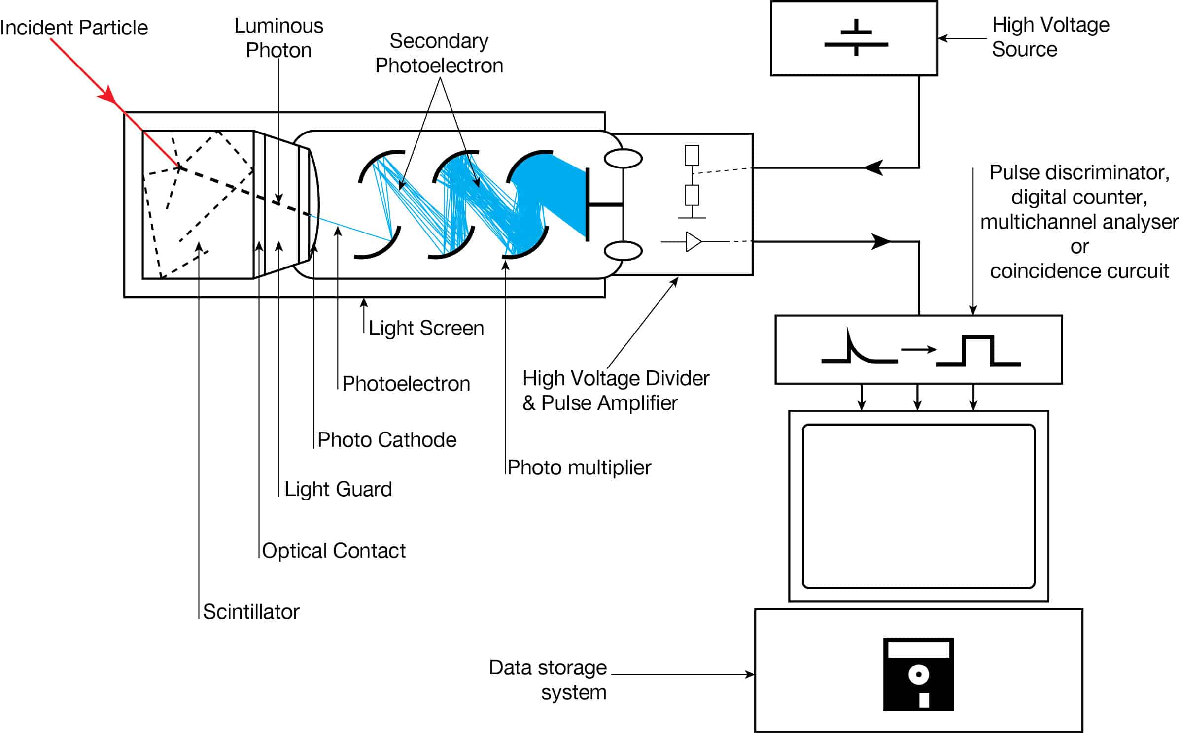
Apparatus with a scintillating crystal, photomultiplier, and data acquisition components. Source: wikipedia.org License CC BY-SA 3.0
Scintillation counters are used to measure radiation in a variety of applications including hand held radiation survey meters, personnel and environmental monitoring for radioactive contamination, medical imaging, radiometric assay, nuclear security and nuclear plant safety. They are widely used because they can be made inexpensively yet with good efficiency, and can measure both the intensity and the energy of incident radiation.
Gamma Rays. High-Z materials are best suited as scintillators for the detection of gamma rays. The most widely used scintillation material is NaI(Tl) (thallium-doped sodium iodide). The iodine provides most of the stopping power in sodium iodide (since it has a high Z = 53). These crystalline scintillators are characterized by high density, high atomic number, and pulse decay times of approximately 1 microsecond (~ 10-6 sec). Scintillation in inorganic crystals is typically slower than in organic ones. They exhibit high efficiency for detection of gamma rays and are capable of handling high count rates. Inorganic crystals can be cut to small sizes and arranged in an array configuration so as to provide position sensitivity. This feature is widely used in medical imaging to detect X-rays or gamma rays. Inorganic scintillators are better at detecting gamma rays and X-rays. This is due to their high density and atomic number which gives a high electron density.
Detection of Gamma Radiation using Semiconductors – HPGe Detectors
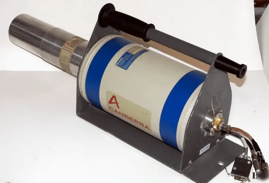
HPGe detector with LN2 cryostat Source: canberra.com
As was written, the study and analysis of gamma ray spectra for scientific and technical use is called gamma spectroscopy, and gamma ray spectrometers are the instruments which observe and collect such data. A gamma ray spectrometer (GRS) is a sophisticated device for measuring the energy distribution of gamma radiation. For the measurement of gamma rays above several hundred keV, there are two detector categories of major importance, inorganic scintillators as NaI(Tl) and semiconductor detectors. If a perfect energy resolution is required, we have to use germanium-based detector, such as the HPGe detector. Germanium-based semiconductor detectors are most commonly used where a very good energy resolution is required, especially for gamma spectroscopy, as well as x-ray spectroscopy. In gamma spectroscopy, germanium is preferred due to its atomic number being much higher than silicon and which increases the probability of gamma ray interaction. Moreover, germanium has lower average energy necessary to create an electron-hole pair, which is 3.6 eV for silicon and 2.9 eV for germanium. This also provides the latter a better resolution in energy. The FWHM (full width at half maximum) for germanium detectors is a function of energy. For a 1.3 MeV photon, the FWHM is 2.1 keV, which is very low.
Robert Reed Burn, Introduction to Nuclear Reactor Operation, 1988.
U.S. Department of Energy, Nuclear Physics and Reactor Theory. DOE Fundamentals Handbook, Volume 1 and 2. January 1993.
Paul Reuss, Neutron Physics. EDP Sciences, 2008. ISBN: 978-2759800414.
See also:
Detectors
We hope, this article, Detection of Gamma Radiation – Detector of Gamma Rays, helps you. If so, give us a like in the sidebar. Main purpose of this website is to help the public to learn some interesting and important information about radiation and dosimeters.
Detection of X-rays is very specific, because high-energy photons interact differently with matter. Detection of X-Rays – Detector of X-Rays
Detection of X-rays is very specific, because high-energy photons interact differently with matter. High-energy photons can travel thousands of feet in air and can easily pass through various materials. Moreover, high-energy photons can ionize atoms indirectly and directly (despite they are electrically neutral) through the photoelectric effect and the Compton effect. But secondary (indirect) ionization is much more significant.
In order to describe principles of detection of high-energy photons, we have to understand the interaction of radiation with matter. Each type particle interacts in a different way, therefore we must describe interactions of high-energy photons (radiation as a flow of these rays) separately.
Although a large number of possible interactions are known, there are three key interaction mechanisms with matter. The strength of these interactions depends on the energy of the X-rays and the elemental composition of the material, but not much on chemical properties, since the X-ray photon energy is much higher than chemical binding energies. The photoelectric absorbtion dominates at low-energies of X-rays, while Compton scattering dominates at higher energies.
The photon is completely absorbed in photoelectric effect, while only partial energy is deposited in any given Compton scattering. The probability of photoelectric absorption (dominates at lower X-rays energies) per unit mass is approximately proportional to:
τ(photoelectric) = constant x ZN/E3.5
where Z is the atomic number, the exponent n varies between 4 and 5. E is the energy of the incident photon. The probability of Compton scattering per one interaction with an atom increases linearly with atomic number Z, because it depends on the number of electrons, which are available for scattering in the target atom.
Detectors of X-Rays
Detectors may be also categorized according to sensitive materials and methods that can be utilized to make a measurement:
Gamma rays have very little trouble in penetrating the metal walls of the chamber. Therefore, ionization chambers may be used to detect gamma radiation and X-rays collectively known as photons, and for this the windowless tube is used. Ionization chambers have a good uniform response to radiation over a wide range of energies and are the preferred means of measuring high levels of gamma radiation. Some problems are caused by the fact, that alpha particles are more ionising than beta particles and than gamma rays, so more current is produced in the ionization chamber region by alpha than beta and gamma. Gamma rays deposit significantly lower amount of energy to the detector than other particles.
Detection of X-Rays using Geiger Counter
Detector of Ionizing Radiation – Geiger Tube
Geiger counter can detect ionizing radiation such as alpha and beta particles, neutrons, X-rays and gamma rays using the ionization effect produced in a Geiger–Müller tube, which gives its name to the instrument. The voltage of detector is adjusted so that the conditions correspond to the Geiger-Mueller region.
The high amplification factor of the Geiger counter is the major advantage over the ionization chamber. Geiger counter is therefore a much more sensitive device than other chambers. It is often used in the detection of low-level gamma rays and beta particles for this reason.
Windowless type
Gamma rays have very little trouble in penetrating the metal walls of the chamber. Therefore, Geiger counters may be used to detect gamma radiation and X-rays (thin-walled tubes) collectively known as photons, and for this the windowless tube is used.
A thick walled tube is used for gamma radiation detection above energies of about 25 KeV, this type generally has an overall wall thickness of about 1-2 mm of chrome steel.
A thin walled tube is used for low energy photons (X-rays or gamma rays) and high energy beta particles. The transition from thin walled to thick walled design takes place at the 300–400 keV energy levels. Above these levels thick walled designs are used, and beneath these levels the direct gas ionisation effect is predominant.
Detection of X-rays using Scintillation Counter
Apparatus with a scintillating crystal, photomultiplier, and data acquisition components. Source: wikipedia.org License CC BY-SA 3.0
Scintillation counters are used to measure radiation in a variety of applications including hand held radiation survey meters, personnel and environmental monitoring for radioactive contamination, medical imaging, radiometric assay, nuclear security and nuclear plant safety. They are widely used because they can be made inexpensively yet with good efficiency, and can measure both the intensity and the energy of incident radiation.
X-Rays.High-Z materials are best suited as scintillators for the detection of gamma rays. The most widely used scintillation material is NaI(Tl) (thallium-doped sodium iodide). The iodine provides most of the stopping power in sodium iodide (since it has a high Z = 53). These crystalline scintillators are characterized by high density, high atomic number, and pulse decay times of approximately 1 microsecond (~ 10-6 sec). Scintillation in inorganic crystals is typically slower than in organic ones. They exhibit high efficiency for detection of gamma rays and are capable of handling high count rates. Inorganic crystals can be cut to small sizes and arranged in an array configuration so as to provide position sensitivity. This feature is widely used in medical imaging to detect X-rays or gamma rays. Inorganic scintillators are better at detecting gamma rays and X-rays. This is due to their high density and atomic number which gives a high electron density.
Detection of X-Rays using Semiconductors – HPGe Detectors
HPGe detector with LN2 cryostat Source: canberra.com
As was written, the study and analysis of gamma ray spectra for scientific and technical use is called gamma spectroscopy, and gamma ray spectrometers are the instruments which observe and collect such data. A gamma ray spectrometer (GRS) is a sophisticated device for measuring the energy distribution of gamma radiation. For the measurement of gamma rays above several hundred keV, there are two detector categories of major importance, inorganic scintillators as NaI(Tl) and semiconductor detectors. If a perfect energy resolution is required, we have to use germanium-based detector, such as the HPGe detector. Germanium-based semiconductor detectors are most commonly used where a very good energy resolution is required, especially for gamma spectroscopy, as well as x-ray spectroscopy. In gamma spectroscopy, germanium is preferred due to its atomic number being much higher than silicon and which increases the probability of gamma ray interaction. Moreover, germanium has lower average energy necessary to create an electron-hole pair, which is 3.6 eV for silicon and 2.9 eV for germanium. This also provides the latter a better resolution in energy. The FWHM (full width at half maximum) for germanium detectors is a function of energy. For a 1.3 MeV photon, the FWHM is 2.1 keV, which is very low.
Robert Reed Burn, Introduction to Nuclear Reactor Operation, 1988.
U.S. Department of Energy, Nuclear Physics and Reactor Theory. DOE Fundamentals Handbook, Volume 1 and 2. January 1993.
Paul Reuss, Neutron Physics. EDP Sciences, 2008. ISBN: 978-2759800414.
See also:
Detectors
We hope, this article, Detection of X-Rays – Detector of X-Rays, helps you. If so, give us a like in the sidebar. Main purpose of this website is to help the public to learn some interesting and important information about radiation and dosimeters.
A Cherenkov detector is a particle detector, which is based on detection of Cherenkov radiation (visible light or UV photons). Cherenkov counters are used mainly for the particle identification. Radiation Dosimetry
A Cherenkov detector is a particle detector, which is based on detection of Cherenkov radiation (visible light or UV photons). In contrast to a scintillation counter the light production is instantaneous. Typical scintillator detectors have a decay time measured in microseconds while Cherenkov radiation is nearly instantaneous and with fast pulse processing equipment can be measured in picoseconds. Cherenkov counters are used mainly for the particle identification, i.e. for the determination of particle masses. Cherenkov counters contain two main elements:
a radiator through which the charged particle passes,
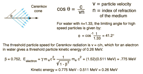
The Cherenkov radiation, which is produced in the radiator, is electromagnetic radiation emitted when a charged particle (such as an electron) moves through a dielectric medium faster than the phase velocity of light in that medium. Particles exceeding the phase velocity of light result in polarization along the axis of motion creating a dipole field. When this field collapses, an electromagnetic pulse (Cherenkov radiation) is emitted in the forward direction. It is similar to the bow wave produced by a boat travelling faster than the speed of water waves. Cherenkov radiation occurs only if the particle’s speed is higher than the phase velocity of light in the material. Even at high energies the energy lost by Cherenkov radiation is much less than that by the other mechanisms (collisions, bremsstrahlung). It is named after Soviet physicist Pavel Alekseyevich Cherenkov, who shared the Nobel Prize in physics in 1958 with Ilya Frank and Igor Tamm for the discovery of Cherenkov radiation, made in 1934.
Source: hyperphysics.phy-astr.gsu.edu
Cherenkov radiation is commonly produced in dielectric materials through Compton electrons or pair-production electrons and positrons. The intensity of light produced by this process is much less than that of luminescence (the basis for scintillation detector operation) requiring more sensitive optical photon detection equipment such as low light photo-multiplier tubes (PMT). Cherenkov counters may be classified as either imaging or threshold types, depending on whether they do or do not make use of Cherenkov angle (θ) information. In the simple case of a threshold detector the mass-dependent threshold energy allows the discrimination between a lighter particle (which does radiate) and a heavier particle (which does not radiate) of the same energy or momentum. Imaging counters may be used to track particles as well as identify them. Although devices using Cherenkov radiation are often thought of as particle identification (PID) detectors, in practice, they are widely used over a much broader range of applications; including:
RICH. Practical multi-track Ring-Imaging Cherenkov detectors (generically called RICH counters) are a more recent development. In a RICH detector, a cone of Cherenkov light is produced when a high speed charged particle traverses a suitable medium, often called radiator. This light cone is detected on a position sensitive planar photon detector, which allows reconstructing a ring or disc, the radius of which is a measure for the Cherenkov emission angle. For example, the LHCb experiment on the Large Hadron Collider uses two RICH detectors for differentiating between pions and kaons.
Super-Kamiokande. Super-Kamiokande is an underground neutrino observatory, which uses large water Cherenkov counters to detect high-energy neutrinos to search for proton decay, study solar and atmospheric neutrinos, and keep watch for supernovae in the Milky Way Galaxy. It consists of a cylindrical stainless steel tank about 40 m (131 ft) in height and diameter holding 50,000 tons of ultrapure water. Mounted on an inside superstructure are about 13,000 photomultiplier tubes that detect light from Cherenkov radiation.
Robert Reed Burn, Introduction to Nuclear Reactor Operation, 1988.
U.S. Department of Energy, Nuclear Physics and Reactor Theory. DOE Fundamentals Handbook, Volume 1 and 2. January 1993.
Paul Reuss, Neutron Physics. EDP Sciences, 2008. ISBN: 978-2759800414.
See also:
Radiation Detection
We hope, this article, Cherenkov Detector, helps you. If so, give us a like in the sidebar. Main purpose of this website is to help the public to learn some interesting and important information about radiation and dosimeters.
Types of Cherenkov Detectors – RICH – Super-Kamiokande. Cherenkov detectors may be classified as either imaging or threshold types, depending on whether they do or do not make use of Cherenkov angle (θ) information. Radiation Dosimetry
Cherenkov detectors may be classified as either imaging or threshold types, depending on whether they do or do not make use of Cherenkov angle (θ) information. In the simple case of a threshold detector the mass-dependent threshold energy allows the discrimination between a lighter particle (which does radiate) and a heavier particle (which does not radiate) of the same energy or momentum. Imaging counters may be used to track particles as well as identify them. Although devices using Cherenkov radiation are often thought of as particle identification (PID) detectors, in practice, they are widely used over a much broader range of applications; including:
RICH. Practical multi-track Ring-Imaging Cherenkov detectors (generically called RICH counters) are a more recent development. In a RICH detector, a cone of Cherenkov light is produced when a high speed charged particle traverses a suitable medium, often called radiator. This light cone is detected on a position sensitive planar photon detector, which allows reconstructing a ring or disc, the radius of which is a measure for the Cherenkov emission angle. For example, the LHCb experiment on the Large Hadron Collider uses two RICH detectors for differentiating between pions and kaons.
Super-Kamiokande. Super-Kamiokande is an underground neutrino observatory, which uses large water Cherenkov counters to detect high-energy neutrinos to search for proton decay, study solar and atmospheric neutrinos, and keep watch for supernovae in the Milky Way Galaxy. It consists of a cylindrical stainless steel tank about 40 m (131 ft) in height and diameter holding 50,000 tons of ultrapure water. Mounted on an inside superstructure are about 13,000 photomultiplier tubes that detect light from Cherenkov radiation.
Robert Reed Burn, Introduction to Nuclear Reactor Operation, 1988.
U.S. Department of Energy, Nuclear Physics and Reactor Theory. DOE Fundamentals Handbook, Volume 1 and 2. January 1993.
Paul Reuss, Neutron Physics. EDP Sciences, 2008. ISBN: 978-2759800414.
See also:
Cherenkov Detectors
We hope, this article, Type of Cherenkov Detectors – RICH – Super-Kamiokande, helps you. If so, give us a like in the sidebar. Main purpose of this website is to help the public to learn some interesting and important information about radiation and dosimeters.
Cherenkov Detector vs Scintillation Detector. Typical scintillator detectors have a decay time measured in microseconds while Cherenkov radiation is nearly instantaneous. Radiation Dosimetry
Cherenkov Detectors
A Cherenkov detector is a particle detector, which is based on detection of Cherenkov radiation (visible light or UV photons). In contrast to a scintillation counter the light production is instantaneous. Typical scintillator detectors have a decay time measured in microseconds while Cherenkov radiation is nearly instantaneous and with fast pulse processing equipment can be measured in picoseconds. Cherenkov counters are used mainly for the particle identification, i.e. for the determination of particle masses. Cherenkov counters contain two main elements:
a radiator through which the charged particle passes,
The Cherenkov radiation, which is produced in the radiator, is electromagnetic radiation emitted when a charged particle (such as an electron) moves through a dielectric medium faster than the phase velocity of light in that medium. Particles exceeding the phase velocity of light result in polarization along the axis of motion creating a dipole field. When this field collapses, an electromagnetic pulse (Cherenkov radiation) is emitted in the forward direction.
Cherenkov radiation is commonly produced in dielectric materials through Compton electrons or pair-production electrons and positrons. The intensity of light produced by this process is much less than that of luminescence (the basis for scintillation detector operation) requiring more sensitive optical photon detection equipment such as low light photo-multiplier tubes (PMT). Cherenkov counters may be classified as either imaging or threshold types, depending on whether they do or do not make use of Cherenkov angle (θ) information. In the simple case of a threshold detector the mass-dependent threshold energy allows the discrimination between a lighter particle (which does radiate) and a heavier particle (which does not radiate) of the same energy or momentum. Imaging counters may be used to track particles as well as identify them. Although devices using Cherenkov radiation are often thought of as particle identification (PID) detectors, in practice, they are widely used over a much broader range of applications; including:
A scintillation counter or scintillation detector is a radiation detector which uses the effect known as scintillation. Scintillation is a flash of light produced in a transparent material by the passage of a particle (an electron, an alpha particle, an ion, or a high-energy photon). Scintillation occurs in the scintillator, which is a key part of a scintillation detector. In general, a scintillation detector consists of:
Scintillator. A scintillator generates photons in response to incident radiation.
Photodetector. A sensitive photodetector (usually a photomultiplier tube (PMT), a charge-coupled device (CCD) camera, or a photodiode), which converts the light to an electrical signal and electronics to process this signal.
The basic principle of operation involves the radiation reacting with a scintillator, which produces a series of flashes of varying intensity. The intensity of the flashes is proportional to the energy of the radiation. This feature is very important. These counters are suited to measure the energy of gamma radiation (gamma spectroscopy) and, therefore, can be used to identify gamma emitting isotopes.
Robert Reed Burn, Introduction to Nuclear Reactor Operation, 1988.
U.S. Department of Energy, Nuclear Physics and Reactor Theory. DOE Fundamentals Handbook, Volume 1 and 2. January 1993.
Paul Reuss, Neutron Physics. EDP Sciences, 2008. ISBN: 978-2759800414.
See also:
Cherenkov Detectors
We hope, this article, Cherenkov Detector vs Scintillation Detector, helps you. If so, give us a like in the sidebar. Main purpose of this website is to help the public to learn some interesting and important information about radiation and dosimeters.
Doping of semiconductors is the intentional introduction of impurities into an intrinsic semiconductor. Dopants that produce the desired controlled changes are classified as either electron acceptors or donors. Radiation Dosimetry
A doped semiconductor, is a semiconductor, that was intentionally doped for the purpose of modulating its electrical, optical and structural properties. In case of semiconductor detectors of ionizing radiation, doping is the intentional introduction of impurities into an intrinsic semiconductor for the purpose of changes in their electrical properties. Therefore, intrinsic semiconductors are also known as pure semiconductors or i-type semiconductors.
The addition of a small percentage of foreign atoms in the regular crystal lattice of silicon or germanium produces dramatic changes in their electrical properties, since these foreign atoms incorporated into the crystal structure of the semiconductor provide free charge carriers (electrons or electron holes) in the semiconductor. In an extrinsic semiconductor it is these foreign dopant atoms in the crystal lattice that mainly provide the charge carriers which carry electric current through the crystal. In general, there are two types of dopant atoms resulting in two types of extrinsic semiconductors. These dopants that produce the desired controlled changes are classified as either electron acceptors or donors and the corresponding doped semiconductors are known as:
n-type Semiconductors.
p-type Semiconductors.
Extrinsic semiconductors are components of many common electrical devices, as well of many detectors of ionizing radiation. For these purpose, a semiconductor diode (devices that allow current in only one direction) usually consists of p-type and n-type semiconductors placed in junction with one another.
Donor Doped Semiconductors
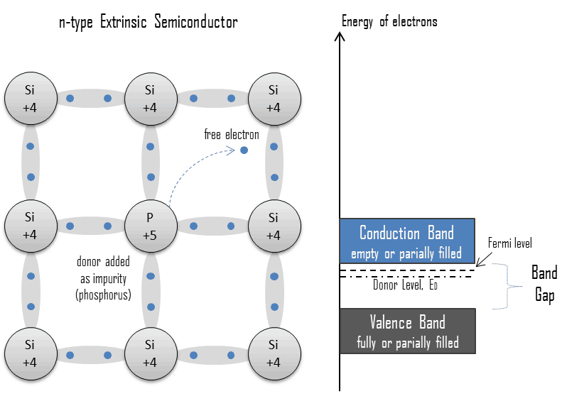
An extrinsic semiconductor which has been doped with electron donor atoms is called an n-type semiconductor, because the majority of charge carriers in the crystal are negative electrons. Since silicon is a tetravalent element, the normal crystal structure contains 4 covalent bonds from four valence electrons. In silicon, the most common dopants are group III and group V elements. Group V elements (pentavalent) have five valence electrons, which allows them to act as a donor. That means, the addition of these pentavalent impurities such as arsenic, antimony or phosphorus contributes free electrons, greatly increasing the conductivity of the intrinsic semiconductor. For example, a silicon crystal doped with boron (group III) creates a p-type semiconductor whereas a crystal doped with phosphorus (group V) results in an n-type semiconductor.
The conduction electrons are completely dominated by the number of donor electrons. Therefore:
The total number of conduction electrons is approximately equal to the number of donor sites, n≈ND.
Charge neutrality of semiconductor material is maintained because excited donor sites balance the conduction electrons. The net result is that the number of conduction electrons is increased, while the number of holes is reduced. The imbalance of the carrier concentration in the respective bands is expressed by the different absolute number of electrons and holes. Electrons are majority carriers, while holes are minority carriers in n-type material.
Acceptor Doped Semiconductors
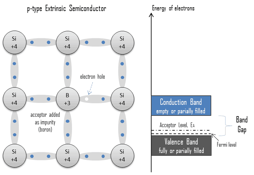
An extrinsic semiconductor which has been doped with electron acceptor atoms is called a p-type semiconductor, because the majority of charge carriers in the crystal are electron holes (positive charge carriers). The pure semiconductor silicon is a tetravalent element, the normal crystal structure contains 4 covalent bonds from four valence electrons. In silicon, the most common dopants are group III and group V elements. Group III elements (trivalent) all contain three valence electrons, causing them to function as acceptors when used to dope silicon. When an acceptor atom replaces a tetravalent silicon atom in the crystal, a vacant state (an electron hole) is created. An electron hole (often simply called a hole) is the lack of an electron at a position where one could exist in an atom or atomic lattice. It is one of the two types of charge carriers that are responsible for creating electric current in semiconducting materials. These positively charged holes can move from atom to atom in semiconducting materials as electrons leave their positions. The addition of trivalent impurities such as boron, aluminum or gallium to an intrinsic semiconductor creates these positive electron holes in the structure. For example, a silicon crystal doped with boron (group III) creates a p-type semiconductor whereas a crystal doped with phosphorus (group V) results in an n-type semiconductor.
The number of electron holes are completely dominated by the number of acceptor sites. Therefore:
The total number of holes is approximately equal to the number of donor sites, p ≈ NA.
Charge neutrality of this semiconductor material is also maintained. The net result is that the number of electron holes is increased, while the number of conduction electrons is reduced. The imbalance of the carrier concentration in the respective bands is expressed by the different absolute number of electrons and holes. Electron holes are majority carriers, while electrons are minority carriers in p-type material.
Robert Reed Burn, Introduction to Nuclear Reactor Operation, 1988.
U.S. Department of Energy, Nuclear Physics and Reactor Theory. DOE Fundamentals Handbook, Volume 1 and 2. January 1993.
Paul Reuss, Neutron Physics. EDP Sciences, 2008. ISBN: 978-2759800414.
See also:
Types of Semiconductors
We hope, this article, Doping of Semiconductors, helps you. If so, give us a like in the sidebar. Main purpose of this website is to help the public to learn some interesting and important information about radiation and dosimeters.
Highly doped semiconductors behave metallically and these semiconductors are known as degenerate (very highly doped) semiconductors. Radiation Dosimetry
An extrinsic semiconductor, or doped semiconductor, is a semiconductor, that was intentionally doped for the purpose of modulating its electrical, optical and structural properties. In case of semiconductor detectors of ionizing radiation, doping is the intentional introduction of impurities into an intrinsic semiconductor for the purpose of changes in their electrical properties. Therefore, intrinsic semiconductors are also known as pure semiconductors or i-type semiconductors.
The addition of a small percentage of foreign atoms in the regular crystal lattice of silicon or germanium produces dramatic changes in their electrical properties, since these foreign atoms incorporated into the crystal structure of the semiconductor provide free charge carriers (electrons or electron holes) in the semiconductor. In an extrinsic semiconductor it is these foreign dopant atoms in the crystal lattice that mainly provide the charge carriers which carry electric current through the crystal. In general, there are two types of dopant atoms resulting in two types of extrinsic semiconductors. These dopants that produce the desired controlled changes are classified as either electron acceptors or donors and the corresponding doped semiconductors are known as:
n-type Semiconductors.
p-type Semiconductors.
Degenerate Semiconductors
It must be noted, increased doping leads to increased conductivity due to the higher concentration of charge carriers. Highly doped semiconductors behave metallically and these semiconductors are known as degenerate (very highly doped) semiconductors. Degenerate semiconductors are often used in integrated circuits as a replacement for metal. Often superscript plus and minus symbols are used to denote relative doping concentration in semiconductors. For example, n+ denotes an n-type semiconductor with a high doping concentration. Similarly, p− would indicate a very lightly doped p-type material.
Robert Reed Burn, Introduction to Nuclear Reactor Operation, 1988.
U.S. Department of Energy, Nuclear Physics and Reactor Theory. DOE Fundamentals Handbook, Volume 1 and 2. January 1993.
Paul Reuss, Neutron Physics. EDP Sciences, 2008. ISBN: 978-2759800414.
See also:
Types of Semiconductors
We hope, this article, Degenerate Semiconductor, helps you. If so, give us a like in the sidebar. Main purpose of this website is to help the public to learn some interesting and important information about radiation and dosimeters.