X-ray dosimetry is very specific, because high-energy photons interact differently with matter. High-energy photons can travel thousands of feet in air and can easily pass through various materials. Moreover, high-energy photons can ionize atoms indirectly and directly (despite they are electrically neutral) through the photoelectric effect and the Compton effect. But secondary (indirect) ionization is much more significant.
Detectors of X-Rays
Detectors may be also categorized according to sensitive materials and methods that can be utilized to make a measurement:
Detection of X-Rays using Ionization Chamber
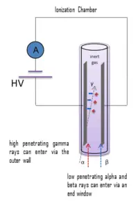
Gamma rays have very little trouble in penetrating the metal walls of the chamber. Therefore, ionization chambers may be used to detect gamma radiation and X-rays collectively known as photons, and for this the windowless tube is used. Ionization chambers have a good uniform response to radiation over a wide range of energies and are the preferred means of measuring high levels of gamma radiation. Some problems are caused by the fact, that alpha particles are more ionising than beta particles and than gamma rays, so more current is produced in the ionization chamber region by alpha than beta and gamma. Gamma rays deposit significantly lower amount of energy to the detector than other particles.
Detection of X-Rays using Geiger Counter
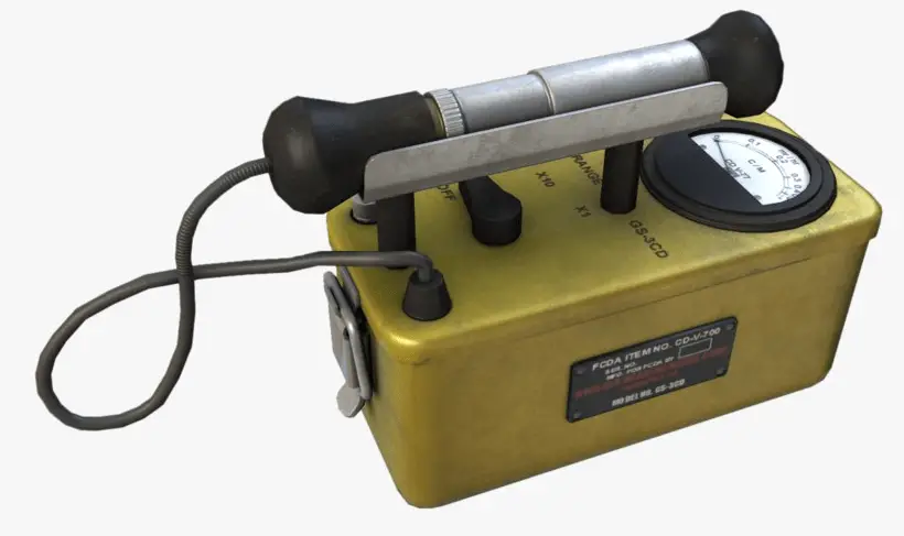
Geiger counter can detect ionizing radiation such as alpha and beta particles, neutrons, X-rays and gamma rays using the ionization effect produced in a Geiger–Müller tube, which gives its name to the instrument. The voltage of detector is adjusted so that the conditions correspond to the Geiger-Mueller region.
The high amplification factor of the Geiger counter is the major advantage over the ionization chamber. Geiger counter is therefore a much more sensitive device than other chambers. It is often used in the detection of low-level gamma rays and beta particles for this reason.
Windowless type
Gamma rays have very little trouble in penetrating the metal walls of the chamber. Therefore, Geiger counters may be used to detect gamma radiation and X-rays (thin-walled tubes) collectively known as photons, and for this the windowless tube is used.
- A thick walled tube is used for gamma radiation detection above energies of about 25 KeV, this type generally has an overall wall thickness of about 1-2 mm of chrome steel.
- A thin walled tube is used for low energy photons (X-rays or gamma rays) and high energy beta particles. The transition from thin walled to thick walled design takes place at the 300–400 keV energy levels. Above these levels thick walled designs are used, and beneath these levels the direct gas ionisation effect is predominant.
Detection of X-rays using Scintillation Counter
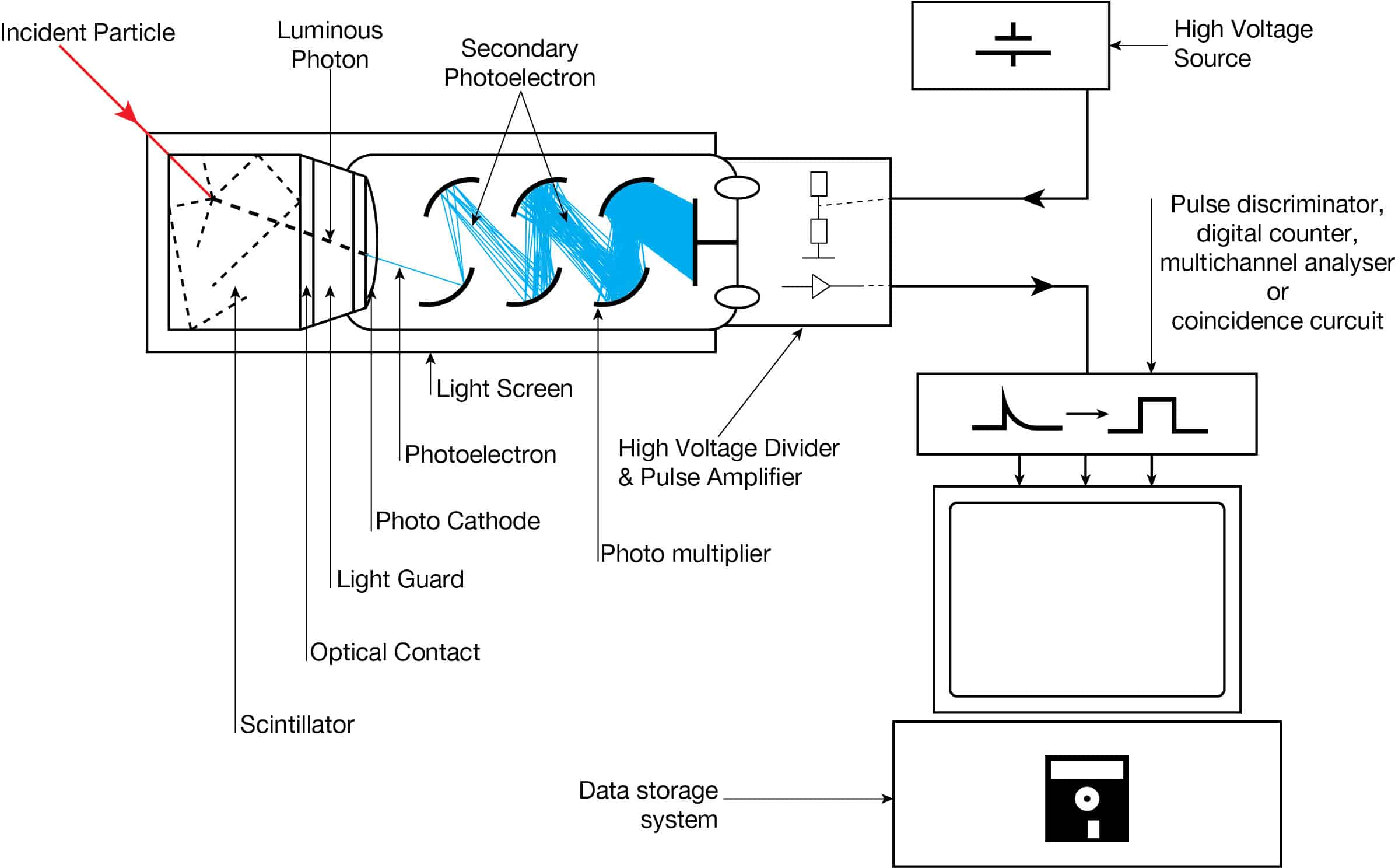
Scintillation counters are used to measure radiation in a variety of applications including hand held radiation survey meters, personnel and environmental monitoring for radioactive contamination, medical imaging, radiometric assay, nuclear security and nuclear plant safety. They are widely used because they can be made inexpensively yet with good efficiency, and can measure both the intensity and the energy of incident radiation.
Scintillation counters can be used to detect alpha, beta, X-rays and gamma radiation. They can be used also for detection of neutrons. For these purposes, different scintillators are used.
- X-Rays. High-Z materials are best suited as scintillators for the detection of gamma rays. The most widely used scintillation material is NaI(Tl) (thallium-doped sodium iodide). The iodine provides most of the stopping power in sodium iodide (since it has a high Z = 53). These crystalline scintillators are characterized by high density, high atomic number, and pulse decay times of approximately 1 microsecond (~ 10-6 sec). Scintillation in inorganic crystals is typically slower than in organic ones. They exhibit high efficiency for detection of gamma rays and are capable of handling high count rates. Inorganic crystals can be cut to small sizes and arranged in an array configuration so as to provide position sensitivity. This feature is widely used in medical imaging to detect X-rays or gamma rays. Inorganic scintillators are better at detecting gamma rays and X-rays. This is due to their high density and atomic number which gives a high electron density.
Detection of X-Rays using Semiconductors – HPGe Detectors
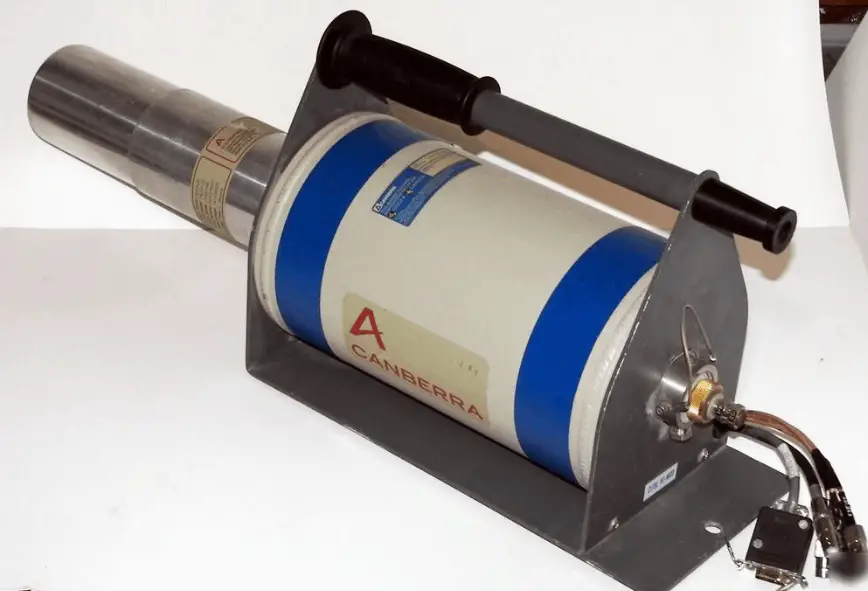
High-purity germanium detectors (HPGe detectors) are the best solution for precise gamma and x-ray spectroscopy.
As was written, the study and analysis of gamma ray spectra for scientific and technical use is called gamma spectroscopy, and gamma ray spectrometers are the instruments which observe and collect such data. A gamma ray spectrometer (GRS) is a sophisticated device for measuring the energy distribution of gamma radiation. For the measurement of gamma rays above several hundred keV, there are two detector categories of major importance, inorganic scintillators as NaI(Tl) and semiconductor detectors. If a perfect energy resolution is required, we have to use germanium-based detector, such as the HPGe detector. Germanium-based semiconductor detectors are most commonly used where a very good energy resolution is required, especially for gamma spectroscopy, as well as x-ray spectroscopy. In gamma spectroscopy, germanium is preferred due to its atomic number being much higher than silicon and which increases the probability of gamma ray interaction. Moreover, germanium has lower average energy necessary to create an electron-hole pair, which is 3.6 eV for silicon and 2.9 eV for germanium. This also provides the latter a better resolution in energy. The FWHM (full width at half maximum) for germanium detectors is a function of energy. For a 1.3 MeV photon, the FWHM is 2.1 keV, which is very low.
EPD – Electronic Personal Dosimeter
An electronic personal dosimeter is modern dosimeter, which can give a continuous readout of cumulative dose and current dose rate, and can warn the person wearing it when a specified dose rate or a cumulative dose is exceeded. EPDs are especially useful in high dose areas where residence time of the wearer is limited due to dose constraints.
Characteristics of EPDs
The electronic personal dosimeter, EPD, is able to display a direct reading of the detected dose or dose rate in real time. Electronic dosimeters may be used as a supplemental dosimeter as well a primary dosimeter. The passive dosimeters and the electronic personal dosimeters are often used together to complement each other. To estimate effective doses, dosimeters must be worn on a position of the body representative of its exposure, typically between the waist and the neck, on the front of the torso, facing the radioactive source. Dosimeters are usually worn on the outside of clothing, around the chest or torso to represent dose to the “whole body”. Dosimeters may also be worn on the extremities or near the eye to measure equivalent dose to these tissues.
The dosimeter can be reset, usually after taking a reading for record purposes, and thereby re-used multiple times. The EPDs have a top mounted display to make them easy to read when they are clipped to your breast pocket. The digital display gives both dose and dose rateinformation usually in mSv and mSv/h. The EPD has a dose rate alarm, and a dose alarm. These alarms are programmable. Different alarms can be set for different activities.
For example:
- dose rate alarm at 100 μSv/h,
- dose alarm: 100 μSv.
If an alarm set point is reached, the relevant display flashes along with a red light, and quite a piercing noise is generated. You can clear the dose rate alarm by retreating to a lower radiation field, but you cannot clear the dose alarm until you get to a EPD reader. EPDs can also give a bleep for every 1 or 10 μSv they register. This gives you an audible indication of the radiation fields. Some EPDs have wireless communication capabilities. EPDs are capable of measuring a wide radiation dose range from routine (μSv) levels to emergency levels (hundreds mSv or units of Sieverts) with high precision, and may display the exposure rate as well as accumulated exposure values. Of the dosimeter technologies, electronic personal dosimeters are generally the most expensive, largest in size, and the most versatile.
We hope, this article, X-Ray Dosimetry – X-Ray Dosimeter, helps you. If so, give us a like in the sidebar. Main purpose of this website is to help the public to learn some interesting and important information about radiation and dosimeters.